


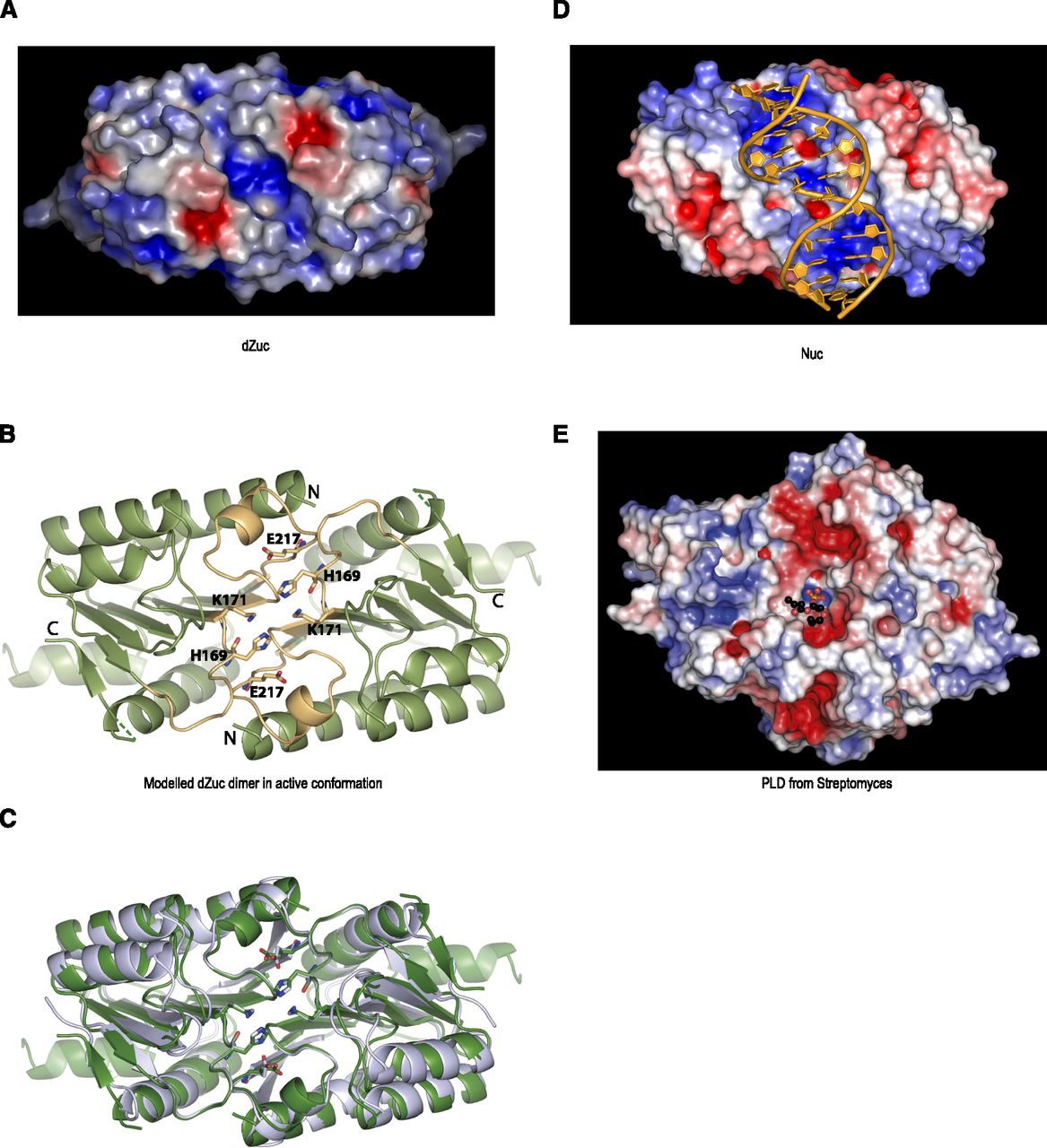
Zucchini dimer probably accepts ssRNA substrates. (A) Surface representation of dZuc dimer colored by electrostatic surface potential (blue, positive; red, negative). The positively charged groove with catalytic residues is too narrow to accommodate a double-stranded nucleic acid. (B) Ribbon representation is shown for the dZuc dimer model that was used in A. It was created based on the related bacterial nuclease Nuc dimer structure, with the active site loops (yellow) and catalytic residues (stick representation) modeled in the catalytically competent state. (C) Overlay of ribbon representation of Nuc dimer (gray) and modeled dZuc dimer (green). (D) Surface representation of Nuc dimer with a double-stranded DNA (orange) modeled into the positively charged groove. (E) Surface representation of a PLD from Streptomyces (PDB 1V0Y) (Leiros et al. 2004) showing electrostatic surface potential (blue, positive; red, negative). The product phospholipid is shown in ball-and-stick representation highlighting the location of the enzyme active site.




