


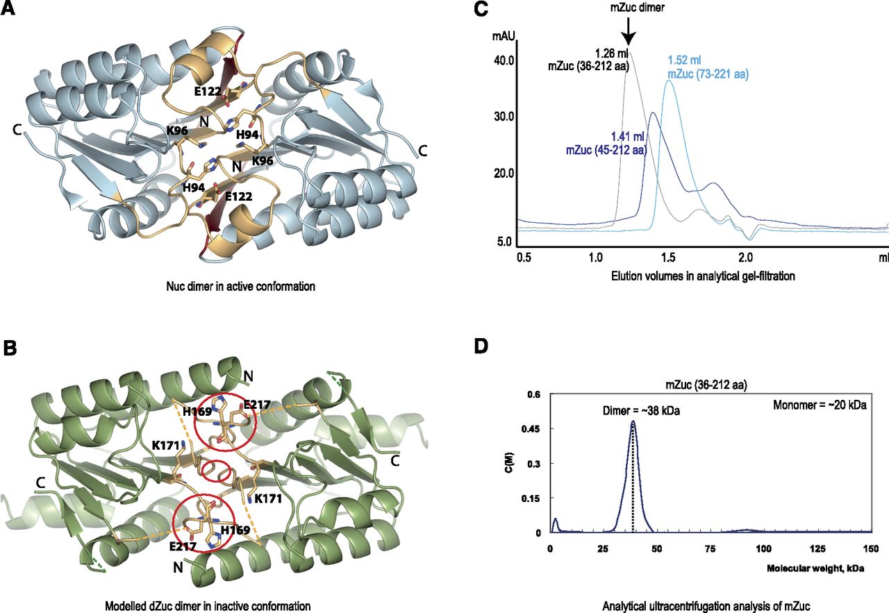
Zucchini forms homodimers. (A) Crystal structure of dimeric Nuc (PDB 1BYR) (Stuckey and Dixon 1999). The N-terminal β-strand (dark red) is required for dimerization. The active site loops (yellow) are in their catalytically competent conformation. (B) dZuc dimer modeled on the related bacterial nuclease Nuc dimer structure. The active site loops (yellow) are misplaced and disordered resulting in clashes on the dimer interface (red circles). (C) Gel-filtration chromatograms for three mouse Zucchini (mZuc) constructs. Dimer formation, as determined by elution volumes (in milliliters), is seen only with the construct mZuc(36–221 aa). (D) Analytical ultracentrifugation analysis of mZuc shows that it sediments as a dimer.




