


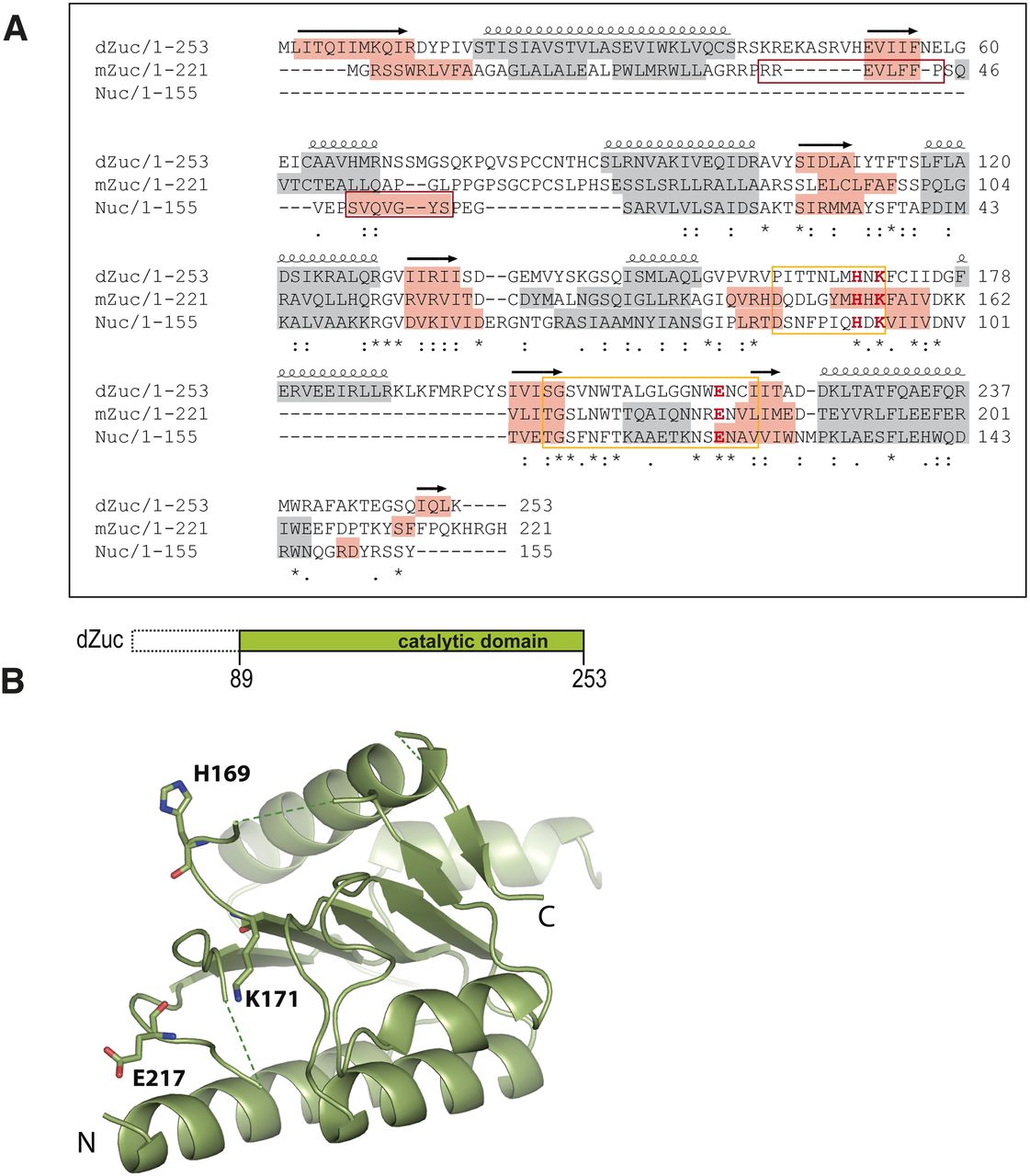
Structure of Drosophila Zucchini. (A) Sequence alignment of Drosophila Zuc (dZuc), mouse Zuc (mZuc), and the bacterial Nuc. Sequence conservation is shown below the alignment. Active site loops that are disordered in the dZuc monomer structure are marked with a yellow box, the catalytic residues shown in the structure figures are highlighted (H, K, E in red), α-helices are shown with gray background, while β-strands are highlighted in orange. The N-terminal β-strand of Nuc (red box) is missing from the Zuc proteins; its role in dimer formation is probably exerted by residues 36–44 in mZuc (note predicted β-strand, red box). (B) The cartoon indicates the dZuc construct used for crystallization. Crystal structure of dZuc is shown in ribbon representation and catalytic residues are shown as sticks. Green dashed lines connect loops with missing density.




