


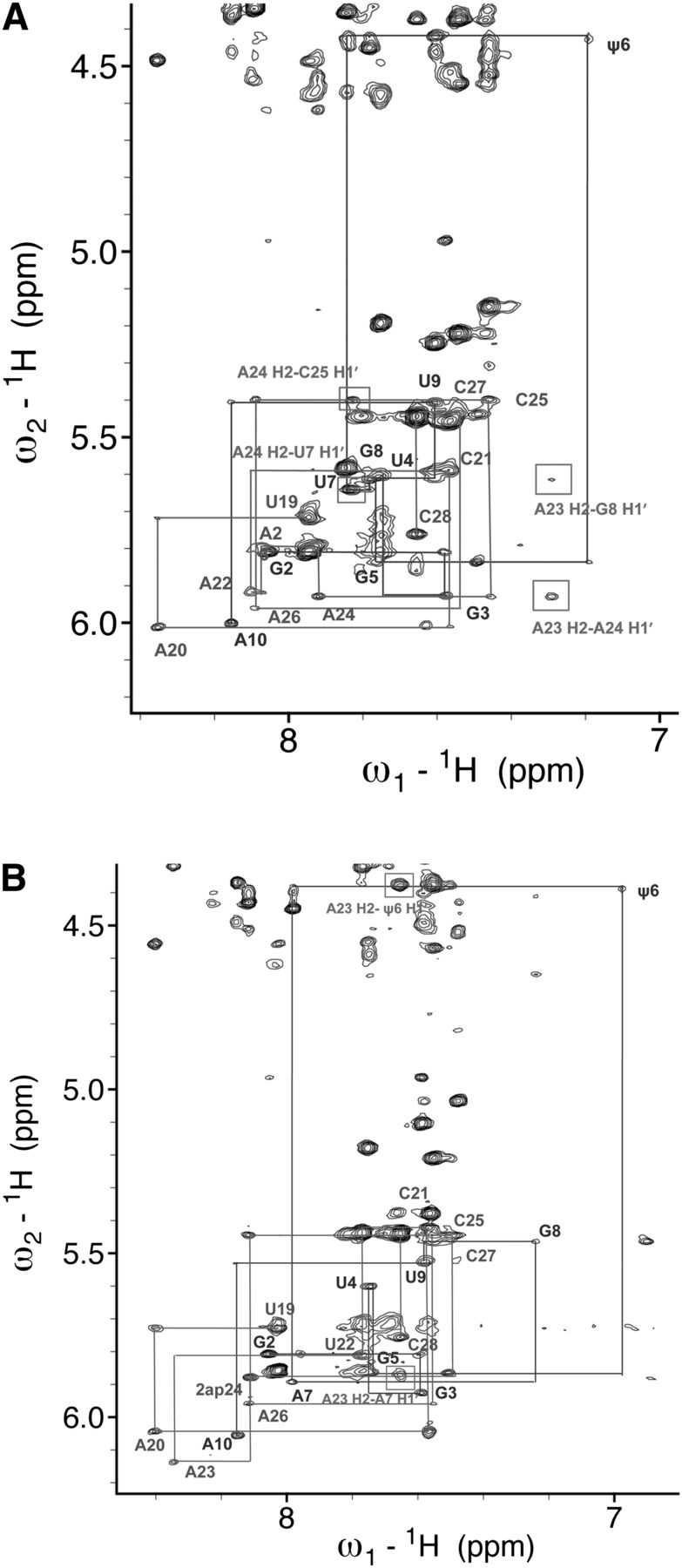
(A) NMR spectrum of the aromatic-anomeric (base-H1′) 1H region of a two-dimensional spectrum of the mutated branch site duplex ψBP2ap. Both spectra shown in A and B were acquired on 0.3 mM RNA, in 99.96% D2O in 50 mM NaCl at 20°C and at 600 MHz with a mixing time of 350 msec. Sequential connectivities between protons H6/H8 and H1′ are marked by lines. Boxed NOEs are those indicating contacts also observed in spectra of ψBP but not in those of uBP (the unmodified duplex in which the branch site A was in an intrahelical conformation) (Newby and Greenbaum 2002a). (B) NMR spectrum of the aromatic-anomeric (base-H1′) 1H region of a two-dimensional spectrum of the mutated branch site duplex (mψBP duplex; 0.3 mM RNA, the same sample conditions as in A). Connectivities between sequential NOEs between protons H6/H8 to H1′ are shown. NOEs involving A24 and A23 H2 protons that confirmed the stacked intrahelical conformation of those residues are marked by rectangles and identified.




