


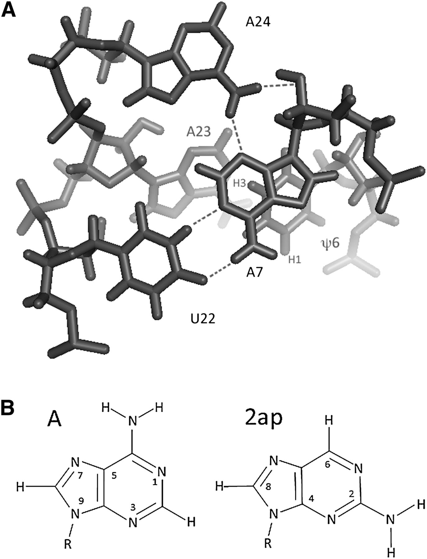
FIGURE 1.
(A) Axial view of the base triple and the adjacent ψ6 and A23 residues in the ψ-modified branch site duplex (ψBP) as seen in the solution structure determined by Newby and Greenbaum (2002a) (1LPW). A7 and U22 participate in a canonical Watson-Crick base pair, while A24, the branch site adenosine residue, adopts an extrahelical conformation and forms a base triple with two hydrogen bonding interactions in the minor group of the A7-U22 pair (with A7 N3 and A7 2′O). (B) Stick structure of adenine and 2-aminopurine (2ap).




