


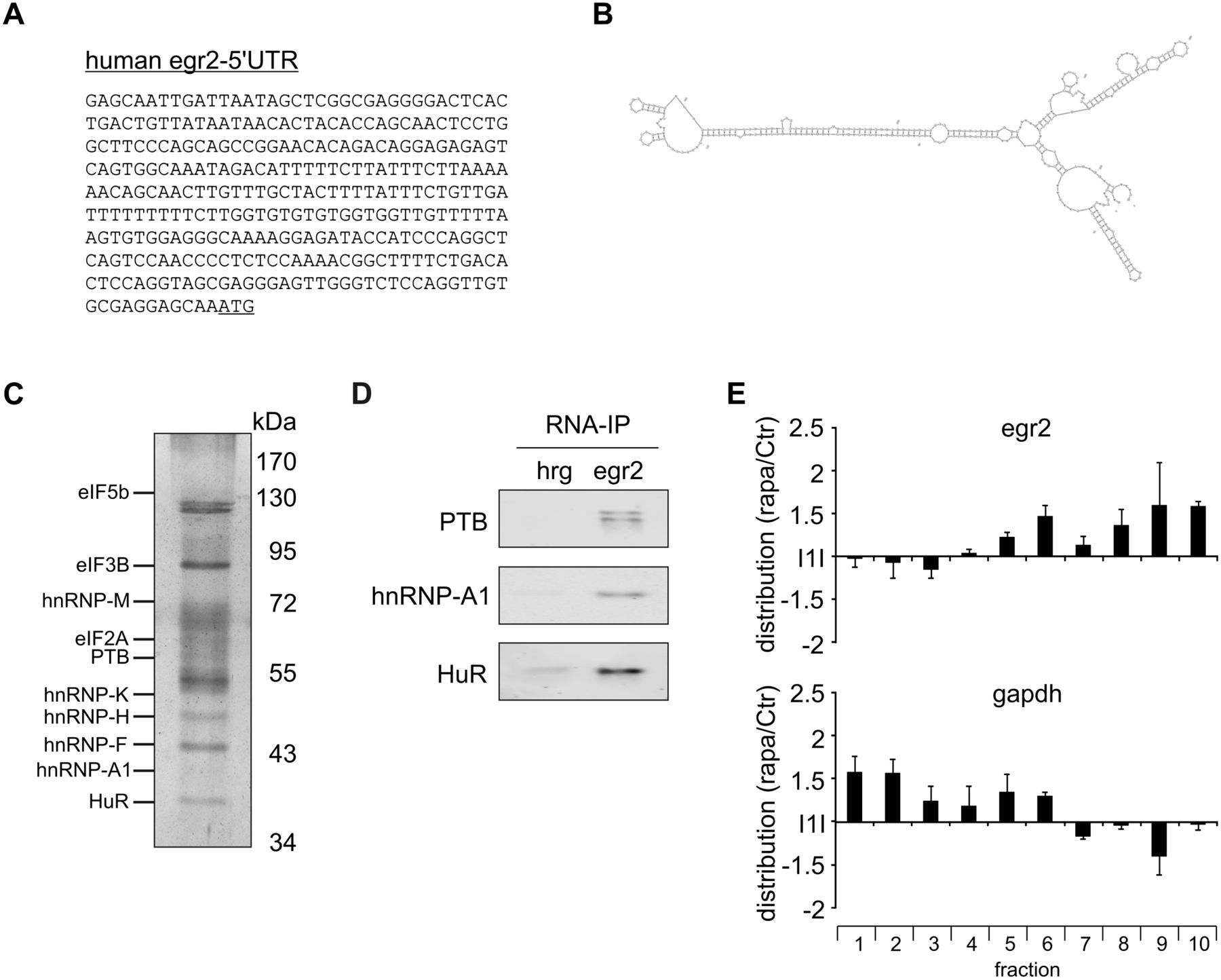
PTB binds to egr2-5′UTR. (A) Sequence of human egr2-5′UTR. (B) Predicted structure of the human egr2-5′UTR using mfold. (C) Lysates of CM-treated MCF7 cells were incubated with in vitro transcribed, biotinylated egr2-5′UTR. After immunoprecipitation using streptavidin agarose beads, bound proteins were separated on SDS-PAGE and visualized using silver staining. Individual proteins were identified by mass spectrometric analysis of gel slices. Positions of identified proteins are shown on the left side, protein molecular weight markers are shown on the right side. (D) Lysates of CM-treated MCF7 cells were incubated with in vitro transcribed, biotinylated egr2-5′UTR or human reverse gapdh (hrg) RNA, before immunoprecipitation of proteins using streptavidin agarose beads. Bound proteins were subjected to Western analysis and probed with the indicated antibodies. Blots are representative for at least three independent experiments. (E) MCF7 cells were treated with rapamycin (100 nM) for 4 h and subjected to polysomal fractionation. RNA from single fractions was isolated and egr2 and gapdh mRNA distribution changes were analyzed using RT-qPCR. Data are presented as means ± SEM (n = 3).




