


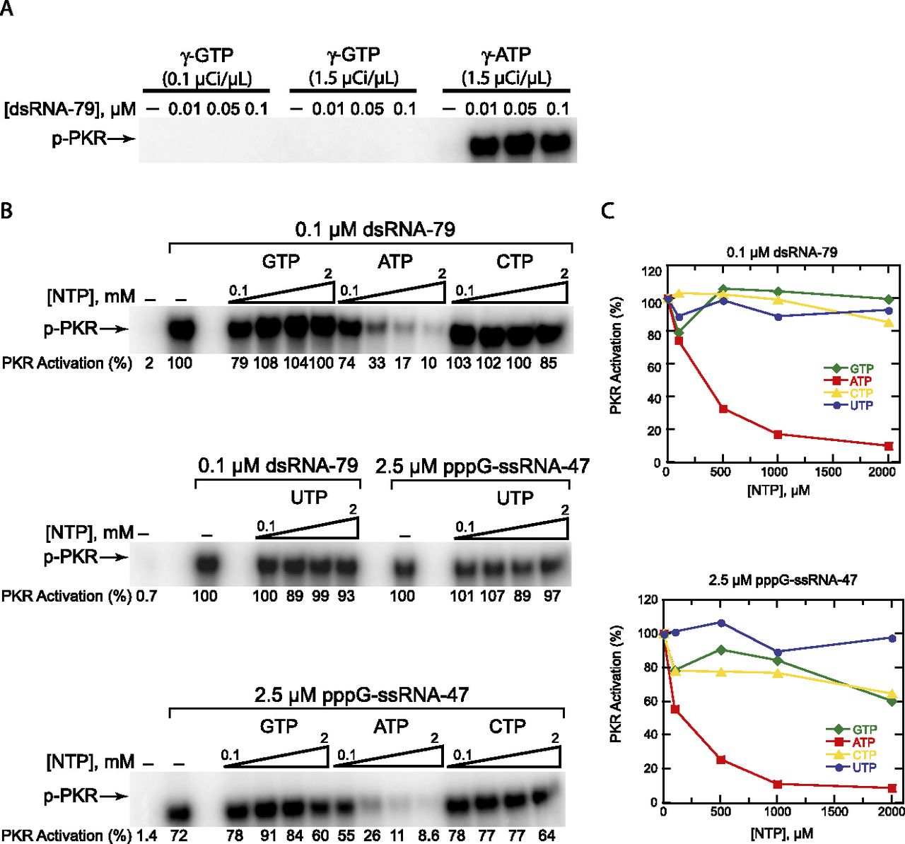
The ATP binding site has high specificity: activation assays. (A) Only ATP supports PKR phosphorylation. Activation of PKR by dsRNA-79 using [γ-32P]GTP or [γ-32P]ATP as the phosphate source. RNA concentrations are provided. In the “γ-GTP” lanes, activation assays were performed as per standard assay conditions (see Materials and Methods) with the following exceptions: 100 μM GTP was used instead of ATP, and 0.1 or 1.5 μCi/μL [γ-32P]GTP was added instead of 1.5 μCi/μL [γ-32P]ATP. In the “γ-ATP” lanes, standard assay conditions were used. (B) NTP-competition assays reveal that only free ATP competes with ATP for activation. PKR activation by dsRNA-79 or pppG-ssRNA-47 was assayed in the presence of increasing concentrations of unlabeled ATP, GTP, CTP, and UTP. The concentrations of dsRNA-79 and pppG-ssRNA-47 were 0.1 and 2.5 μM, respectively. The concentrations of each NTP were 0.1, 0.5, 1, and 2 mM. (All of these are added concentrations and are in the background of 100 μM ATP.) The “no-RNA” and “no-competitor-NTP” lanes are included as negative and positive controls, respectively. Phosphorylation activities were normalized to the “no-competitor-NTP” lane in the middle gel. For both A and B, a 10% SDS-PAGE gel is shown, with the position of phosphorylated PKR (p-PKR) indicated. (C) Graphical representation of phosphorylation activities from panel B.




