


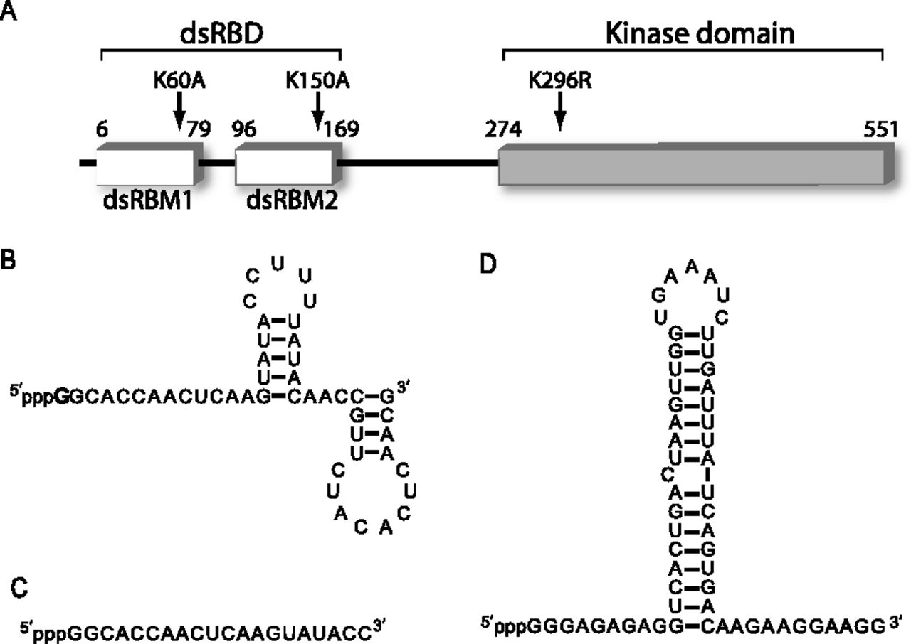
Protein and RNA constructs used in this study. (A) Schematic of PKR primary sequence. The N-terminal dsRNA binding domain (dsRBD, also referred to as P20), composed of two tandem dsRNA-binding motifs (dsRBM 1 and 2), and the catalytic C-terminal kinase domain are indicated. The positions of point mutations used in this study are indicated. The double-mutant PKR (dmPKR) contains both K60A and K150A mutations, and the K296R mutation in the kinase domain renders PKR catalytically inactive. (B) Experimentally determined secondary structure of ssRNA-47 (Nallagatla et al. 2007). The starting nucleotide is indicated in boldface; G is shown, which is typical of transcripts made from WT T7 polymerase, but RNAs were also transcribed containing pppA, pppC, and pppU using mutant T7 polymerase. (C) Sequence of ssRNA-20. Secondary structure prediction via free energy minimization (mFold) indicates that this RNA is essentially completely unstructured. (D) Experimentally determined secondary structure of ss-dsRNA (9,11) (Zheng and Bevilacqua 2004).




