


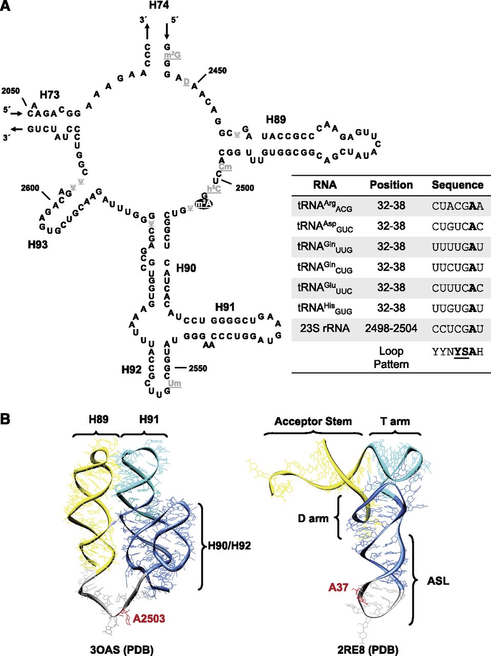
Sequence and structural comparisons of the RlmN substrates. (A) Sequence and secondary structure of the peptidyl transferase region of the E. coli 23S rRNA. Modification m2A at position 2503 is highlighted in black. The remaining post-transcriptional modifications in the region are highlighted in gray. Note location of helices 90–92, which are crucial for recognition by RlmN. In the bottom right box, a short multiple sequence alignment (seven positions in length) of 23S rRNA and the tRNAs carrying m2A37 is used to search for the potential identity determinants recognized by the m2A-synthesizing enzyme. Positions at the consensus pattern are represented according to the IUPAC nucleotide code. Positions 35 and 36 of substrate tRNAs showing a significant conservation pattern were chosen herein for subsequent studies. (B) The three-dimensional structure corresponding to 2455–2580 nucleotides of the Escherichia coli 23S rRNA sequence was extracted from the 3OAS structure stored in the PDB database and shown in ribbon representation (left-hand side) using UCSF Chimera viewer (Pettersen et al. 2004). Helices required for optimal RlmN action (Yan et al. 2010) are depicted in color, and the target nucleoside for the RlmN-mediated methylation is highlighted in red. The three-dimensional, global structure of the Escherichia coli tRNAGlnCUG (extracted from the 2RE8 PDB record) is shown in ribbon representation on the right-hand side. The D, anticodon, and T stem–loops are depicted in color, resembling the secondary structures shown on the left-hand side. The target nucleoside for RlmN-mediated modification is also highlighted in red.




