


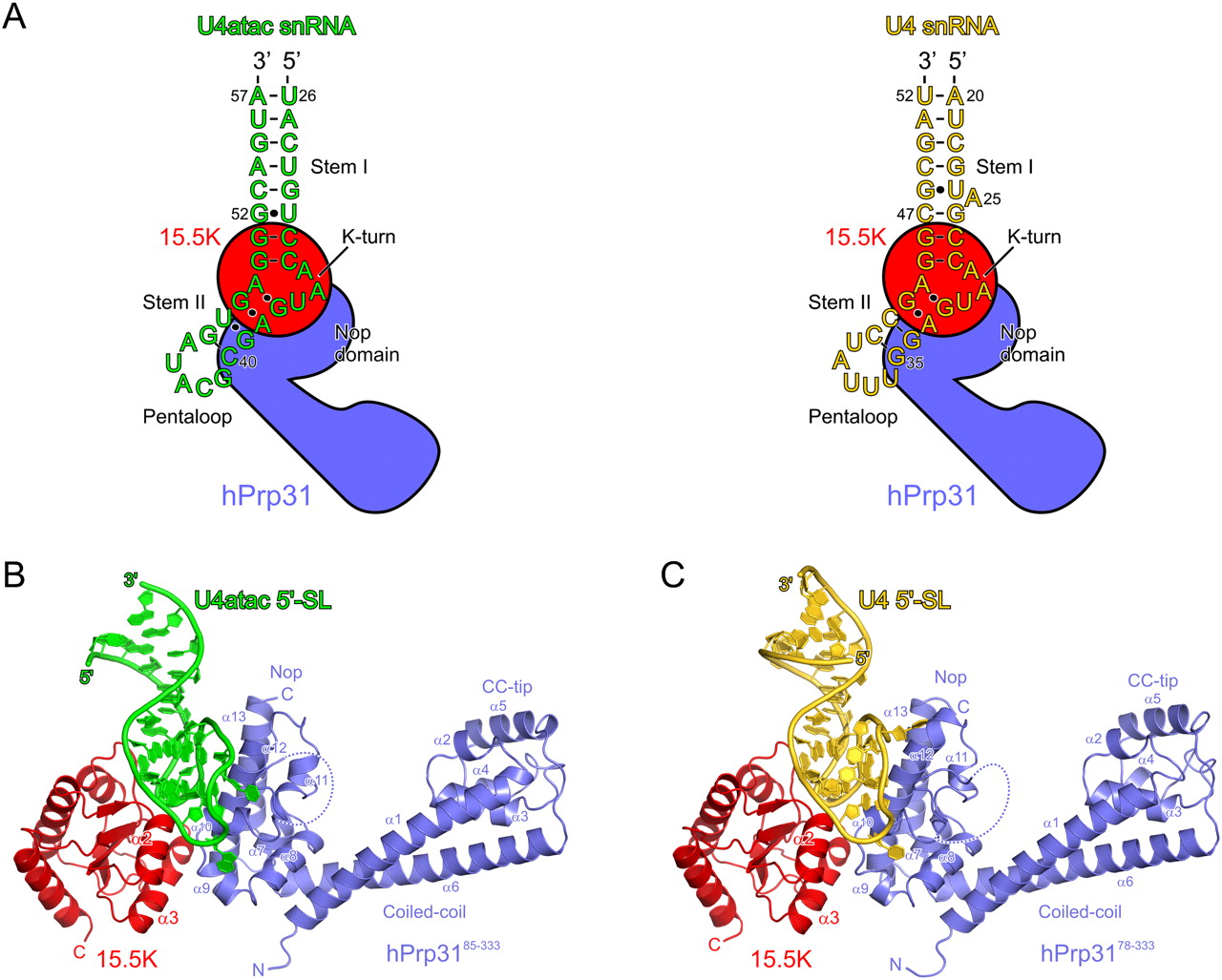
FIGURE 1.
Overall structure of the hPrp3185–333–15.5K–U4atac complex. (A) Schematics of the 5′SL of U4 snRNA and U4atac snRNA. Binding of 15.5K (red) and hPrp31 (steel blue) is indicated schematically. (B) Overview of the hPrp3185–333–15.5K–U4atac 5′SL complex. hPrp3185–333, steel blue; 15.5K, red; U4atac RNA, green. The dashed line in hPrp3185–333 indicates a disordered loop. Secondary structure elements are labeled. (C) The structure of the hPrp3178–333–15.5K–U4 5′SL complex (PDB ID: 2OZB) shown in the same orientation. Coloring as in B, except U4 RNA is shown in gold.




