


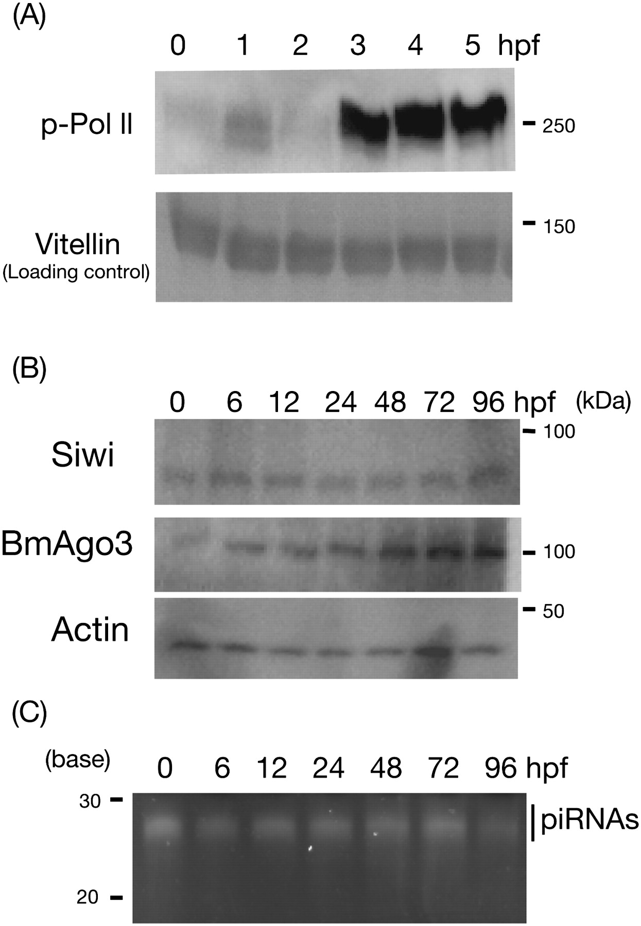
FIGURE 1.
Maternal to zygote transition and maternal deposition of PIWI/piRNA complexes in the silkworm embryos. (A) Proteins from eggs of 0, 1, 2, 3, 4, and 5 hpf were analyzed with anti-phosphorylated RNA polymerase II antibody. Vitellin, a major egg protein served as a loading control. (B) Proteins from eggs of 0, 6, 12, 24, 48, 72, and 96 h post-fertilization (hpf) were analyzed by Western blotting with anti-Siwi, anti-BmAgo3, and anti-actin antibodies. Actin served as a loading control. (C) 10 μg of total RNAs from 0-, 6-, 12-, 24-, 48-, 72-, and 96-hpf eggs were analyzed by urea-containing acrylamide gel. Signals were visualized by SYBRGold staining.




