


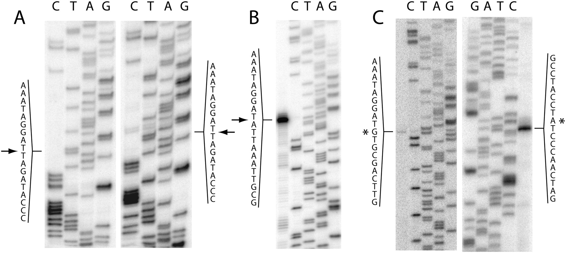
Mapping of intron–exon junctions and the branch site. Sequencing lanes are labeled by the base complementary to the dideoxynucleotide added. (A) Sequencing by reverse transcription of gel-extracted ligated exons; (left panel) Pycnoporellus; (right panel) Grifola. (Arrows) Splicing junctions. (B) Mapping of the 5′ extremity of gel-extracted linear intron molecules generated by in vitro self-splicing of a Pycnoporellus precursor transcript; the latter was used as a template to generate the sequencing lanes at right with a primer located downstream from the intron 5′ extremity. Elongation from the same primer using the excised intron molecules as template generated the strong stop in the lane at left; (arrow) the 5′ splice site. (C) Mapping of the branchpoint and 5′ extremity of gel-extracted lariat intron molecules generated by in vitro self-splicing of a Grifola precursor transcript. (Left panel) Elongation from a primer located downstream from the intron 5′ extremity; the stop (marked by an asterisk) corresponds to the first intron nucleotide; sequencing lanes (at right) were generated by the same primer on a precursor RNA template. (Right panel) elongation from a primer located in the 3′ exon (intron-3′exon branched molecules were used as template); (asterisk) the branch site (elongation stops on the nucleotide immediately 3′ of the branch site); sequencing lanes were generated by the same primer on a precursor RNA template.




