


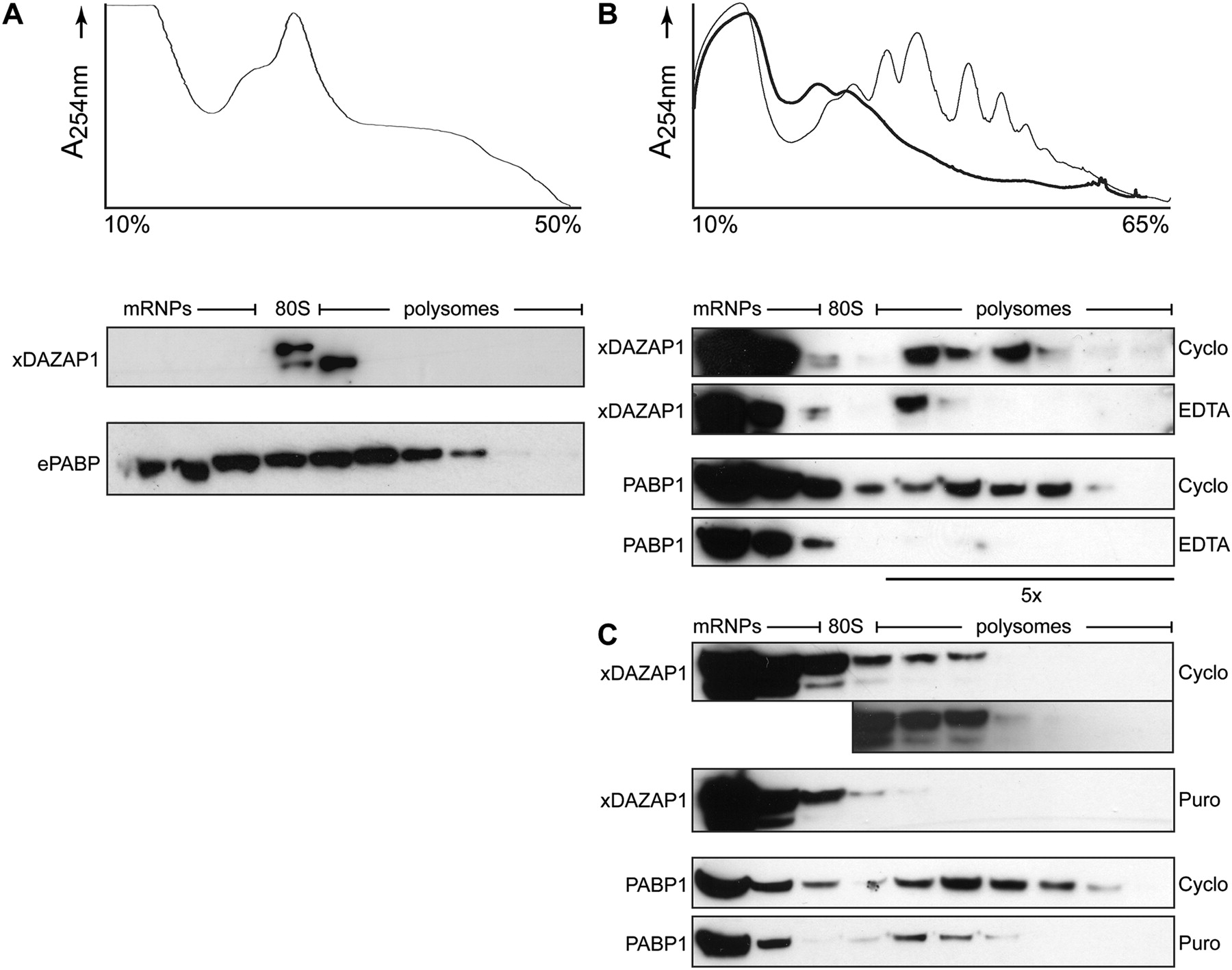
Polysomal association of X. laevis DAZAP1. Stage VI X. laevis oocytes (A) or stage 40–41 embryos (B,C) were subjected to sucrose gradient analysis, and the resulting UV absorbance profiles (254 nm) of cycloheximide-(Cyclo, thin line) or EDTA-treated (thick line) extracts are shown. In stage VI oocytes, polysome peaks are not observed, as only 2% of ribosomes are actively engaged in translation, with the majority of ribosome subunits associating as empty 80S couples. By stage 41, polysome peaks can be clearly discerned. The resulting fractions were probed by Western blot for xDAZAP1 and either ePABP (A) or PABP1 (B,C), depending on developmental stage. EDTA (B) and puromycin (puro; C) treatment both cause the majority of xDAZAP1 and PABP1 to be released from polysomes. In B, five times more protein was loaded into fractions 5–10 to allow enhanced visualization of the association of DAZAP1 and PABP1 with polysomes. The longer exposure of polysomal fractions in C shows that both forms of xDAZAP1 associate with polysomes. The positions of polysomes, the 80S monosome, and mRNP fractions are indicated.




