


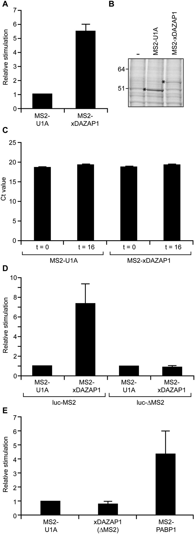
X. laevis DAZAP1 can stimulate translation. (A) Oocytes expressing MS2-xDAZAP1 or MS2-U1A were co-injected with luc-MS2 reporter and β-galactosidase control mRNAs. Luciferase values were corrected for any minor changes in the levels of β-galactosidase and plotted as relative stimulation compared with the control fusion protein MS2-U1A, which is set to 1. (B) SDS-PAGE analysis of 35S-Met–labeled uninjected (−) oocytes or those injected with MS2-U1A or MS2-xDAZAP1 mRNAs. MS2-U1A and MS2-xDAZAP1 contain 21 and 13 methionine residues, respectively. The position of fusion proteins are indicated by asterisks. (C) mRNA was isolated from the oocytes in A either immediately after injection (t = 0) or after 16 h (t = 16), and mRNA levels were determined by qRT-PCR. Mean Ct values are shown. (D) Oocytes expressing MS2-U1A or MS2-xDAZAP1 were injected with β-galactosidase and luciferase mRNAs with (luc-MS2) or without (luc-ΔMS2) MS2-binding sites. (E) luc-MS2 and β-galactosidase mRNAs were injected into oocytes expressing MS2-U1A, ΔMS2-xDAZAP1, or MS2-PABP1, which serves as a positive control. Data in D and E are plotted as described in A.




