


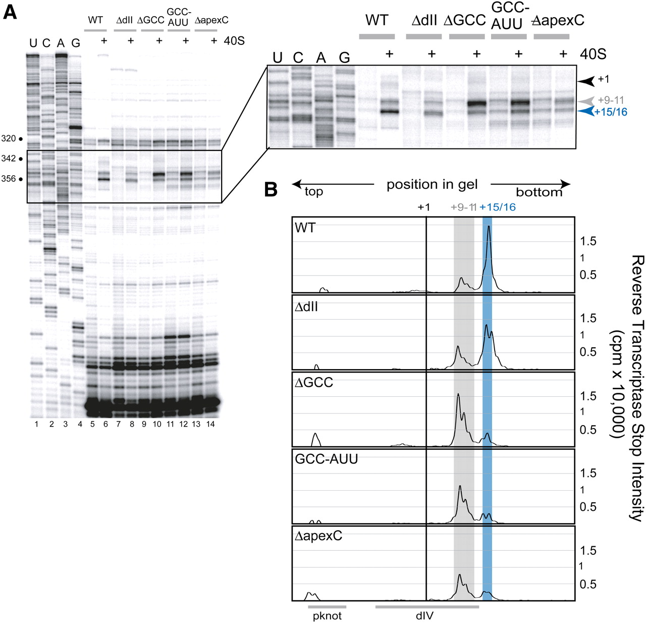
FIGURE 7.
Toeprinting analysis of WT and mutant IRES RNAs. (A) Toeprinting gel of all mutants in the free form and bound to the 40S subunit. The relevant part of this gel is boxed and expanded to the right. The location of the A in the AUG is indicated as the +1 position, and stops at the +9–11 position and at location +15/16 are indicated by arrows to the right of the expanded gel region. (B) Quantitated, background-corrected, and graphed toeprints of IRES•40S from the gel in A. Location +1 (AUG) is indicated by the black line, the +16 toeprint by the blue, and the +9–11 by the gray line. The location of IRES secondary structural regions is shown beneath the graphs.




