


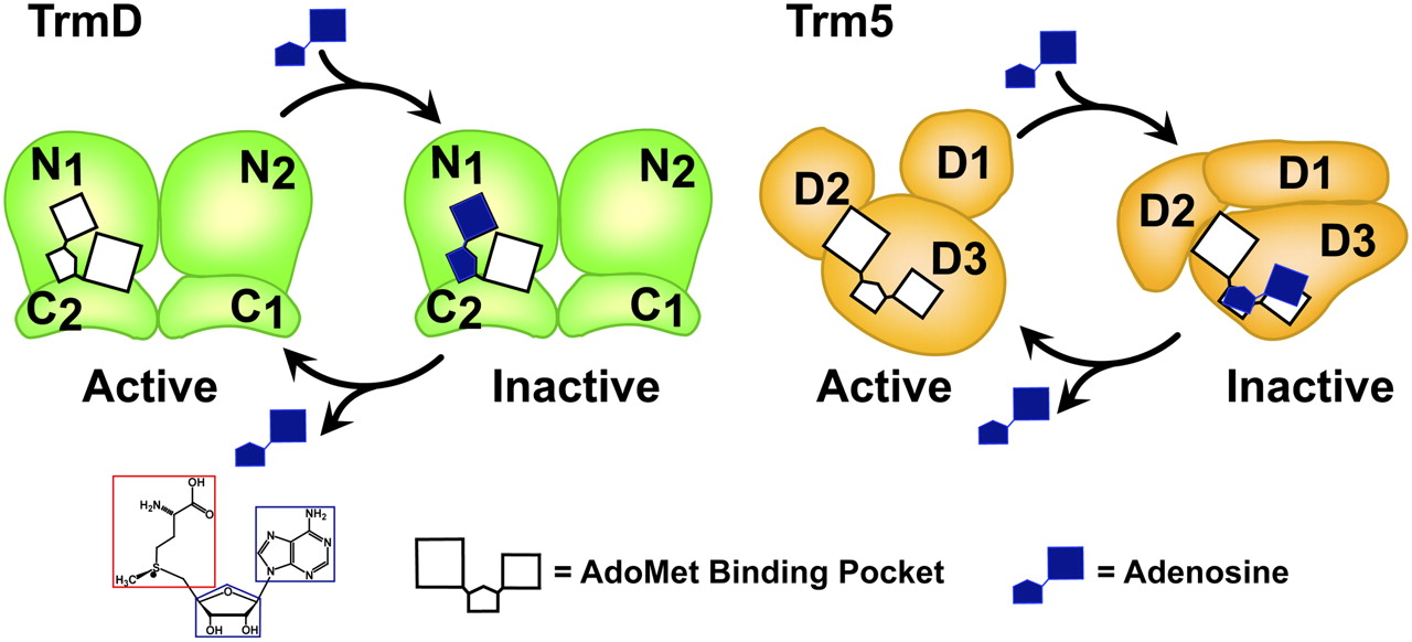
A model of adenosine targeting TrmD and Trm5. In the active state, TrmD and Trm5 each possess an AdoMet-binding site, consisting of an adenosine and a methionine pocket. The AdoMet site is located in a dimer interface of TrmD comprised of the N-terminal domain of monomer 1 (N1) and the C-terminal domain of monomer 2 (C2), whereas it is located between the D2 and D3 domains of the monomeric Trm5. In the adenosine-bound inactive state, TrmD maintains the same structure and binds adenosine in the already-established adenosine pocket, whereas Trm5 shifts its adenosine pocket and binds adenosine in a misfit conformation, leading to structural alterations of the enzyme. In TrmD, the stable binding of adenosine causes delayed release of the ligand, whereas in Trm5 the unstable binding of adenosine can lead to rapid release of the fragment, allowing refolding of the enzyme to its native structure. The chemical structure of AdoMet is depicted below with a blue box showing the adenosine fragment and a red box showing the methionine fragment. The AdoMet-binding site is shown in white, whereas the adenosine ligand is shown in dark blue.




