


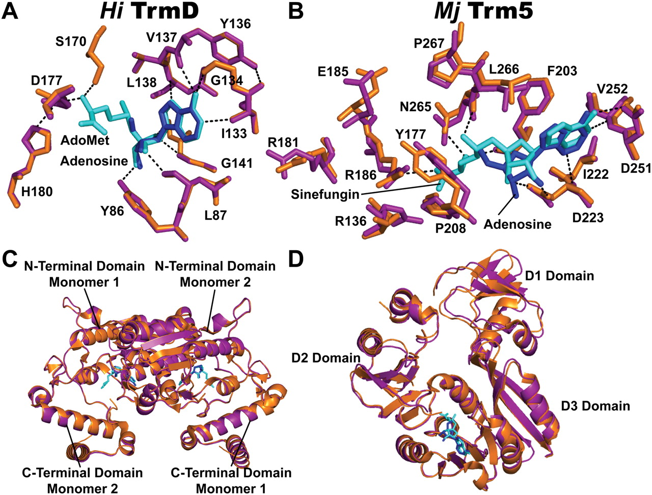
Structural analysis of adenosine-bound TrmD and Trm5 complexes. (A) Superposition of the Cα atoms of adenosine in the adenosine-bound (magenta) complex onto the structure of the AdoMet-bound complex of H. influenzae TrmD (orange, PDB ID: 1UAK). Adenosine and AdoMet are colored in blue and cyan, respectively. Enzyme residues that form the interacting network with AdoMet are depicted, showing H-bonds (dashed lines) with the methyl donor. These residues are in overlapping positions in the two structures. (B) Superposition of the Cα atoms of adenosine in the adenosine-bound (magenta) complex onto the sinefungin-bound complex of M. jannaschii Trm5 (orange, PDB ID: 2YX1). Adenosine and sinefungin are colored in blue and cyan, respectively. Enzyme residues that form the interacting network with sinefungin are depicted, showing H-bonds (dashed lines) with the AdoMet analog. The superposition reveals a shift of adenosine toward the methionine pocket by 0.6 Å with the sugar flexed away from the active site by 1.35 Å. Residues with the most significant shifts are those that contact G37-tRNA in the D2 domain, including Y177 and R136. (C) A ribbon diagram showing the superposition of the overall adenosine-bound and AdoMet-bound structures of H. influenzae TrmD. (D) A ribbon diagram showing the superposition of the overall adenosine-bound and sinefungin-bound structures of M. jannaschii Trm5.




