


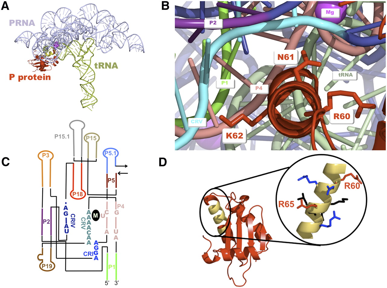
(A) Crystal structure of T. maritima RNase P holoenzyme•tRNA complex (Reiter et al. 2010). (Blue) PRNA; (red) P protein; (green) tRNA. (Yellow) The RNR motif is highlighted on the protein; (magenta ball) the 5′ end of the mature tRNA. (B) Crystal structure of the T. maritima RNase P holoenzyme–tRNA complex (Reiter et al. 2010) with PRNA (dark blue), pre-tRNA (light blue), and P protein (red). The PRNA helices near the RNR motif that comprise the catalytic core of the enzyme are highlighted: P1 (bright green), P2 (purple), P4 (pink), CRV (cyan). The P protein RNR motif residues are displayed as sticks. K62 in the T. maritima corresponds to residue R62 in the B. subtilis holoenzyme. (C) Secondary structure diagram of the B. subtilis PRNA catalytic domain. (M) The general region where metals have been observed to bind in crystal structures of the PRNA (Kazantsev et al. 2009) and RNase P holoenzyme (Reiter et al. 2010). (D) Crystal structure of the B. subtilis RNase P protein (Stams et al. 1998) is shown on the left in red with the RNR motif highlighted in yellow. The RNR motif is enlarged and the residues are color-coded according to their level of conservation (Jovanovic et al. 2002) (red, 100% conservation; blue, 75%–88% conservation; and black, <75% conservation).




