


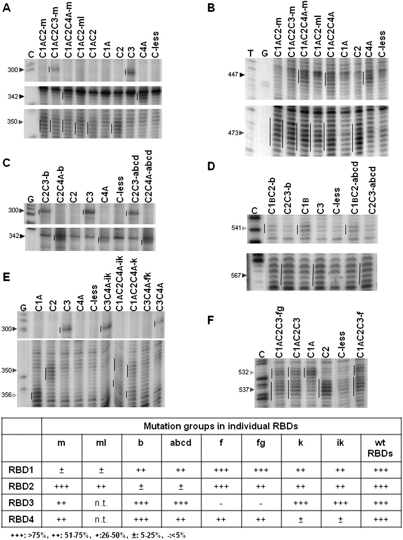
Representative primer extension analyses of directed hydroxyl radical probing experiments to test whether inactivation of one RBD affects the interaction of the other three RBDs with the EMCV and PV IRESs. (A,C,E) Assays of interactions with the 5′-proximal part of the EMCV IRES, using mutants inactivated in RBD1 (panel A), RBD2 (panel C), and RBD4 (panel E). (B,D,F) Assays of interactions with the PV-1 IRES, using mutants inactivated in RBD1 (panel B), RBD2 (panel D), and RBD3 (panel F). In each panel, the vertical line to the left of the reference standard lane highlights the cleavage products generated (in the absence of any inactivating mutations) by Fe(II) conjugated to the particular RBD under examination, and they are repeated in all the other lanes assaying PTB′ derivatives with Fe(II) conjugated to the same RBD. The arrowheads to the left of each panel indicate the reference standard cleavage site position in the IRESs. Lanes C,G,T are from sequencing ladders using the same primer. The cleavage band intensities in these and many other similar experiments were quantitated as described in Materials and Methods, and the cleavage band intensity determined for PTB′ derivatives with inactivating mutations in one RBD and Fe(II)-BABE conjugated to one or more non-mutated RBDs was compared with that found for the corresponding reference standard (WT) with no inactivating mutations (taken as 100%). The consensus of the relative intensity values obtained from many such experiments, covering all three PTB binding sites (two in the EMCV IRES and one in the PV IRES), is tabulated in a semi-quantitative form at the bottom of the figure.




