


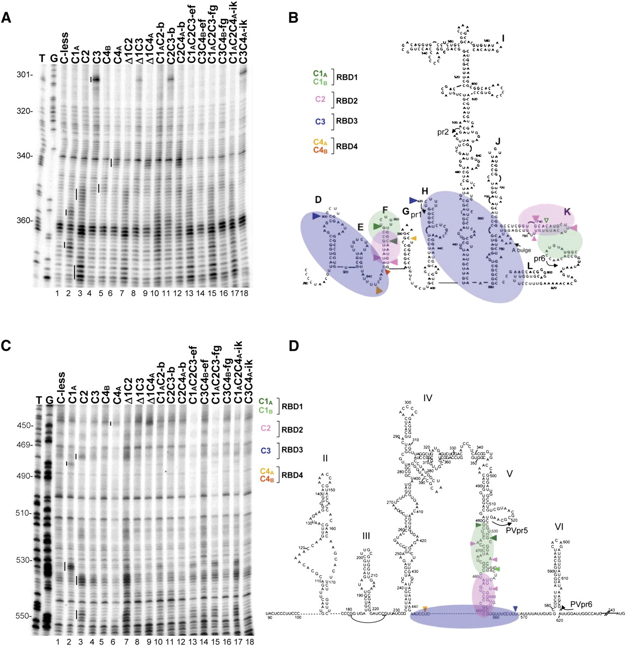
Representative primer extension analyses of directed hydroxyl radical probing of the EMCV and PV IRESs with Fe(II)-BABE PTB′ mutants. (A) Directed hydroxyl radical probing was carried out under standard conditions with the designated Fe(II)-PTB′ derivatives, and the cleavage products were analyzed by primer extension using EMCVpr1 to examine nt 300–375 of the EMCV IRES. Sites of hydroxyl radical cleavages are indicated by vertical lines to the left of each reference standard (control) lane. Lanes T,G are from sequencing ladders using the same primer. (B,D) Schematic representation of the orientation of PTB binding to the EMCV IRES (panel B), adapted from Kafasla et al. (2009), and to the PV-1 IRES (panel D), adapted from Kafasla et al. (2010). RBD1 is in transparent green, pink denotes RBD2, and the RBD3 + 4 didomain is in transparent blue. The same color coding is used to show the sites of cleavage by the designated Fe(II)-PTB′ reference standard derivatives. Cleavages are classified as strong (large filled arrowhead), medium (medium filled arrowhead), or weak (smallest open arrowhead). For clarity, only the cleavages used in the present study as reference standards are shown; for complete cleavage site maps, see Kafasla et al. (2009, 2010). The annealing positions of the primers used for the primer extension analyses in the present study are also shown. (C) Tethered hydroxyl radical probing of PV IRES RNA with the specified Fe(II)-PTB′ mutants, using primer PVpr6 for primer extension analysis of nt 445–555. Cleavage sites are highlighted by vertical lines, as in panel A.




