


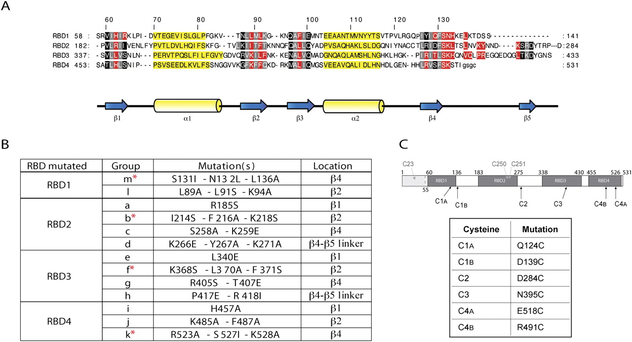
Generation of PTB′ mutants to disrupt the RNA-binding potential of individual RBDs. (A) Sequence alignment of the four RBDs of PTB, adapted from Oberstrass et al. (2005), with the same color coding: amino acid residues interacting with the RNA in red; residues in black and gray are located in the β-sheets, with those in gray pointing toward the hydrophobic core of the RBD; and residues in yellow are in the α-helices. (B) The mutation groups introduced into each RBD to disrupt RNA-binding. Their location in each RBD is given, and the asterisks identify the most effective inactivating mutation group in each RBD. (C) Schematic representation of PTB1 showing the positions of the four RBDs, the first (N-terminal) 54 amino acids that were deleted in PTB′ (gray shading) and the native cysteines of PTB (in gray) which were substituted with serines in PTB′. The positions of the single cysteines introduced into each RBD and used as reference standards are also shown, and the actual amino acid replacements are listed.




