


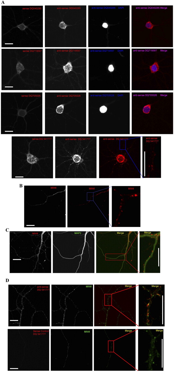
Localization of piRNAs and MIWI in cultured hippocampal neuron. (A) In situ hybridization of DIV21 mouse hippocampal neurons was performed with respective LNA-based sense and anti-sense probes as indicated. The nucleus was stained with DAPI; the merged image shows cytoplasmic localization of piRNAs. Although three piRNAs (DQ540285, DQ719597, and DQ705026) are restricted to the cell soma, piRNA (DQ541777) extended to the neuronal dendrite. In situ hybridization with sense control did not show hybridization signal. A portion of neuron with high-resolution image shown here is marked with a red box in Supplemental Figure 6. Scale bars, 20 μm. (B) Mouse hippocampal neurons at DIV21 were immunostained with an antibody against MIWI. Similar to RNA binding proteins, MIWI also showed a punctate pattern through the neuronal dendrite as well as the cell body. A portion of neuron with high-resolution image shown here is marked with a red box in Supplemental Figure 6. Scale bar, 20 μm. (C) Dendritic localization of piRNA (DQ 541777) was confirmed by immunostaining of mouse hippocampal neurons (DIV21) with a dendritic marker, MAP2. MIWI puncta colocalized with MAP2. A portion of neuron with high-resolution image shown here is marked with a red box in Supplemental Figure 6. Scale bars, 20 μm. (D) Association of piRNA and MIWI was analyzed by in situ hybridization of mouse hippocampal neurons (DIV21) using LNA-based anti-sense or sense probes and immunostained with a MIWI antibody. Both piRNA (DQ541777) and MIWI showed a punctate pattern in the neuronal dendrite. piRNA (DQ541777) and MIWI puncta showed colocalization throughout the neuronal dendrite. In situ hybridization with a sense control did not show any dendritic signal. A portion of neuron with high-resolution image shown here is marked with a red box in Supplemental Figure 6. Scale bars, 20 μm.




