


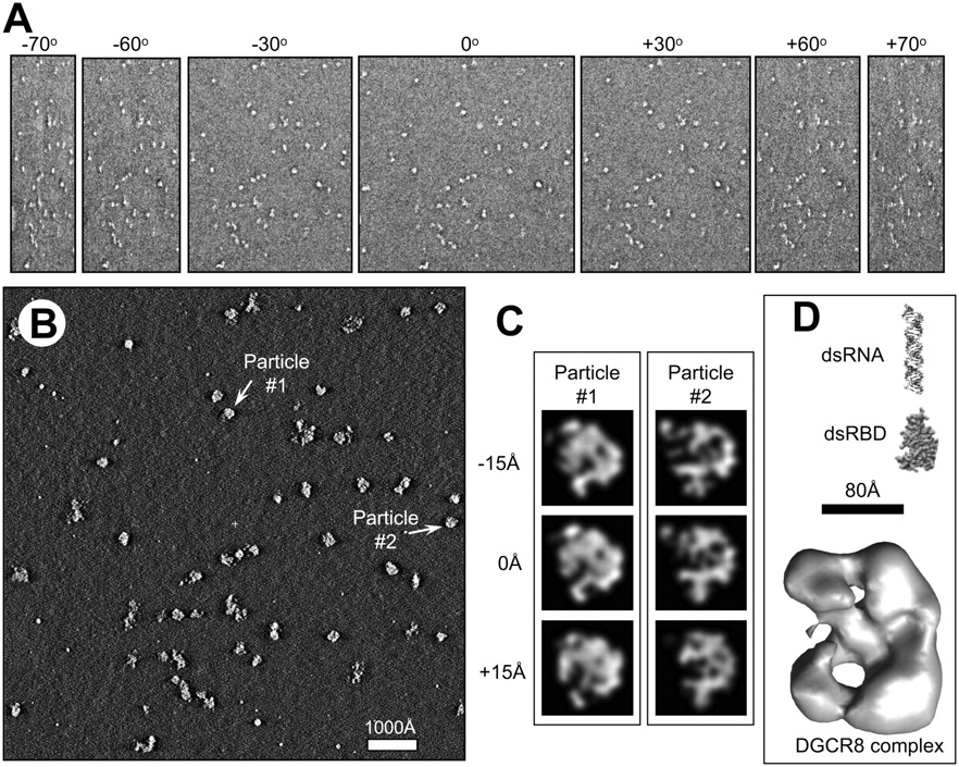
Three-dimensional (3D) structural analysis of the DGCR8–pri-mir-30a complex by electron tomography. (A) Representative tilt images from an electron tomography single tilt series of 141 images, showing the negatively stained DGCR8–pri-mir-30a complex particles imaged from −70° to +70°. Each tilt series was acquired with an angular interval of one degree of tilt between images. (B) A slice through the 3D tomogram reconstructed from the tilt series shown in A after alignment. (C) Illustration of subtomographic volume averaging of individual DGCR8–pri-mir-30a complex particles. Two particles selected from the 3D tomogram as indicated in B were segmented out, low pass filtered, and rotated to create particles suitable for averaging. Shown here are single pixel (7.4 Å) slices at three different z-heights of two particles segmented from the reconstruction in B and aligned in 3D. Particles aligned this way were averaged together to generate an averaged 3D density map of the complex to improve signal/noise ratio and to minimize distortions due to the missing wedge problem intrinsic to electron tomography. (D) Surface representation of the averaged density map of DGCR8–pri-mir-30a complex. As a comparison of sizes and shape, we also show a model of the double-stranded region of miR-30a hairpin and the crystal structure of the dsRBDs of DGCR8 (Sohn et al. 2007).




