


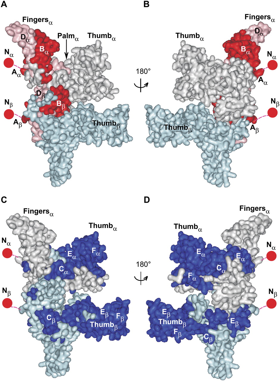
Three-dimensional model of LtrA highlighting regions required for binding of DIVa and intron RNA core regions. (A,B) Front and back views of the model with regions A and B identified as being required for DIVa binding by unigenic evolution analysis (Fig. 3) shown in red. Region D, which may enhance LtrA binding to DIVa by stabilizing the fold of regions A and/or B (see text), is shown in pink. (C,D) Front and back views of the model showing regions of LtrA required for Ll.LtrB-ΔDIVa intron splicing (Fig. 6) highlighted in dark blue. The LtrA subunit modeled on HIV-1 RT p66 (subunit α) is in light gray, and the LtrA subunit modeled on HIV-1 p51 (subunit β) is in light cyan. The model encompasses LtrA amino acid residues 37–538. The N-terminal 36 amino acid residues, which could not be modeled based on HIV-1 RT, are represented as spheres at the N-termini of the monomers (Nα and Nβ, respectively). Abbreviations: (A–F) regions defined by unigenic evolution analyses in Figures 3 and 6, with subscripts α and β referring to the subunit.




