


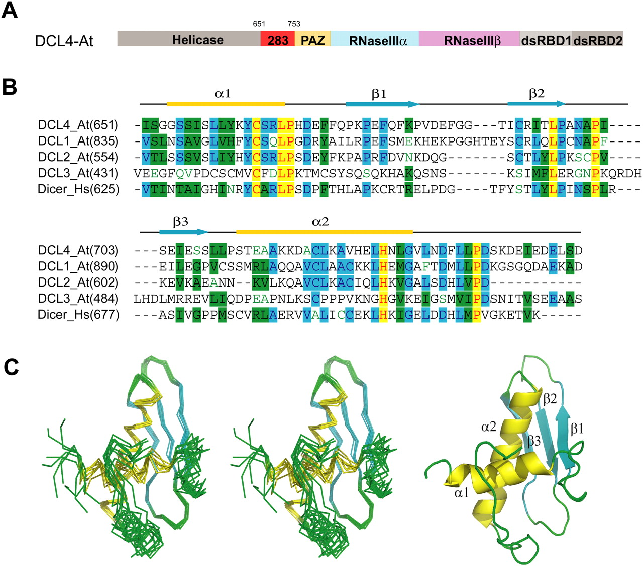
Overall structure of DCL4 DUF283. (A) Domain architecture of Arabidopsis thaliana DCL4. (B) Sequence alignment and secondary structure of Dicer DUF283. The aligned sequences (Swiss Protein ID) are in the order of DCL4_At, DCL1_At, DCL2_At, DCL3_At, and Dicer_Hs. The secondary structure diagram for DCL4_At is shown on top. The α-helices are colored in yellow, β-strands are colored in cyan. Conserved residues are shaded in cyan (80% similarity) and green (60% similarity), whereas essentially invariant residues are shaded in yellow. (C) Stereo view of the ensemble of eight lowest energy NMR structures of the DCL4 DUF283 domain (residues 651–752) and Ribbon representation of the DCL4 DUF283 domain.




