


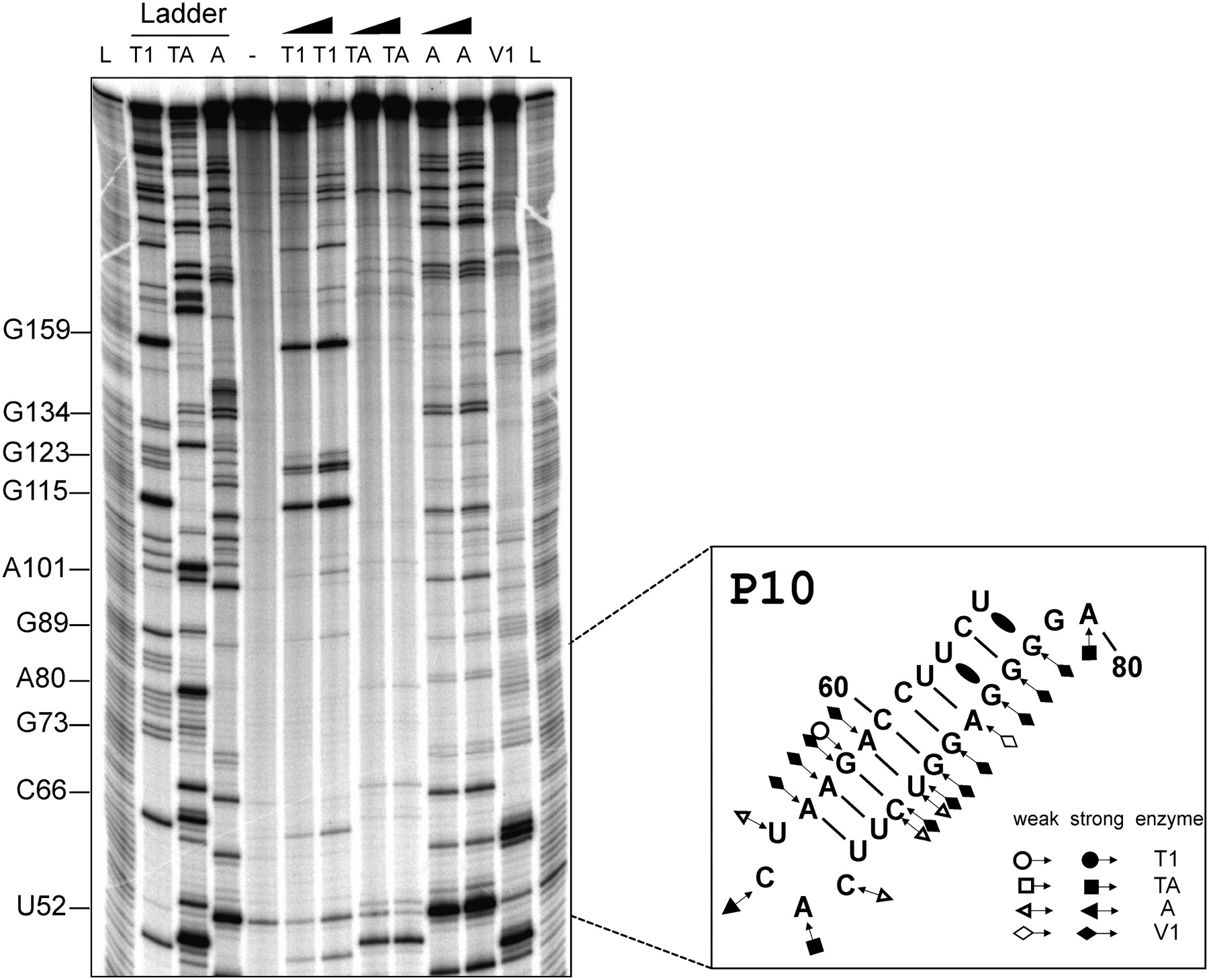
Typical autoradiogram of an 8% PAGE gel performed for the enzymatic probing of a 5′-end labeled PLMVd transcripts of minus polarity. The lanes labeled L on both sides are identical ladders obtained by alkaline hydrolysis of PLMVd. Lanes 2, 3, and 4 contain PLMVd transcripts hydrolyzed by RNase T1, RNase TA, and RNase A, respectively, under denaturing conditions in order to provide guanosine, adenosine, and cytosine/uridine ladders. Lane 5 is an unhydrolyzed RNA sample. The remaining lanes contained samples obtained from hydrolyzes performed using two dilutions of RNase T1 (lanes 6,7), RNase TA (lanes 8,9), RNase A (lanes 10,11), and one dilution of RNase V1 (lane 12). The inset on the right is a schematic compilation of the RNase probing of the P10 stem–loop.




