


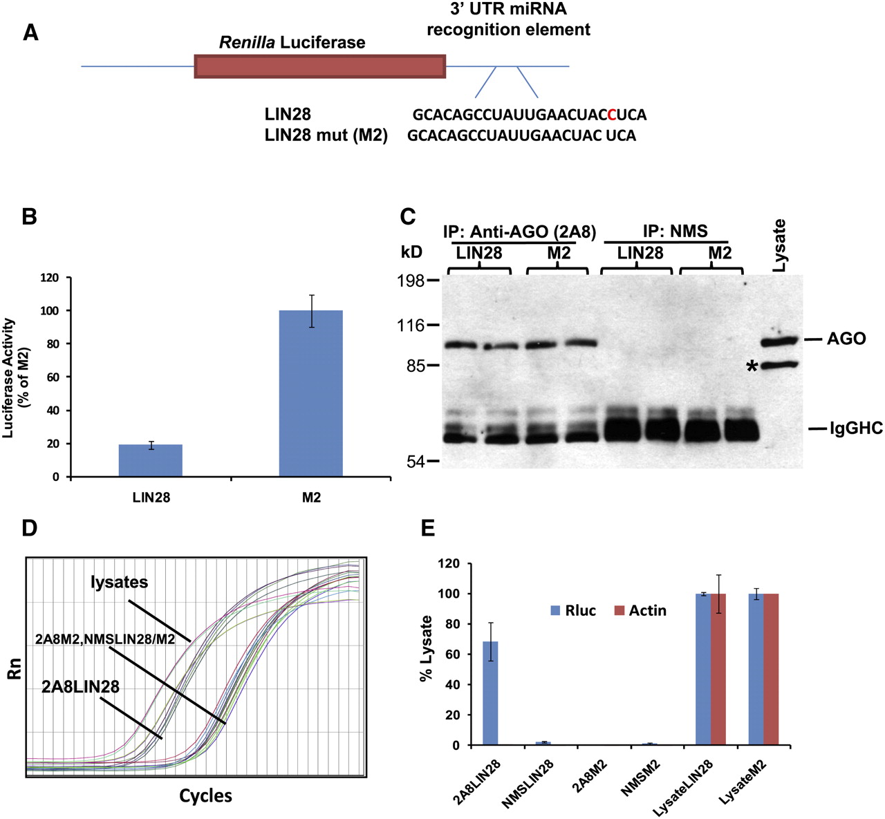
RIP-Chip was validated in H4 cells using positive miRNA target control and closely matched negative controls with transfected Renilla luciferase (Rluc) reporter plasmids. (A) Let-7 miRNA recognition element (MRE) of LIN28 or its closely related mutated sequence (designated “M2”) in the 3′-untranslated region (UTR) of the Rluc gene as previously described (Kiriakidou et al. 2004). (B) Dual luciferase activity assay shows that LIN28 reporter activity was decreased as expected by endogenous Let-7 miRNA. (C) Western blots analysis of co-IPed products using anti-AGO antibody. As expected, AGO proteins were co-IPed with anti-AGO (2A8), but not with nonimmunized mouse serum (NMS). Co-IPs were performed on cells transfected with reporter plasmid Rluc-LIN28 or Rluc-M2. An asterisk (*) shows a band that is Radixin, a protein recognized on Western blots, but not via co-IP by anti-AGO 2A8. (D) Although AGO protein was co-IPed regardless of the transfected plasmids, RT-qPCR detected Rluc mRNA only in the anti-AGO co-IP from the cell lysates transfected with Rluc-LIN28. (E) Quantification of qPCR shows that Rluc mRNA was highly enriched only in co-IPed RNAs from cells transfected with Rluc-LIN28, demonstrating a highly specific association of Rluc mRNA with AGO proteins via miRNA binding sites.




