


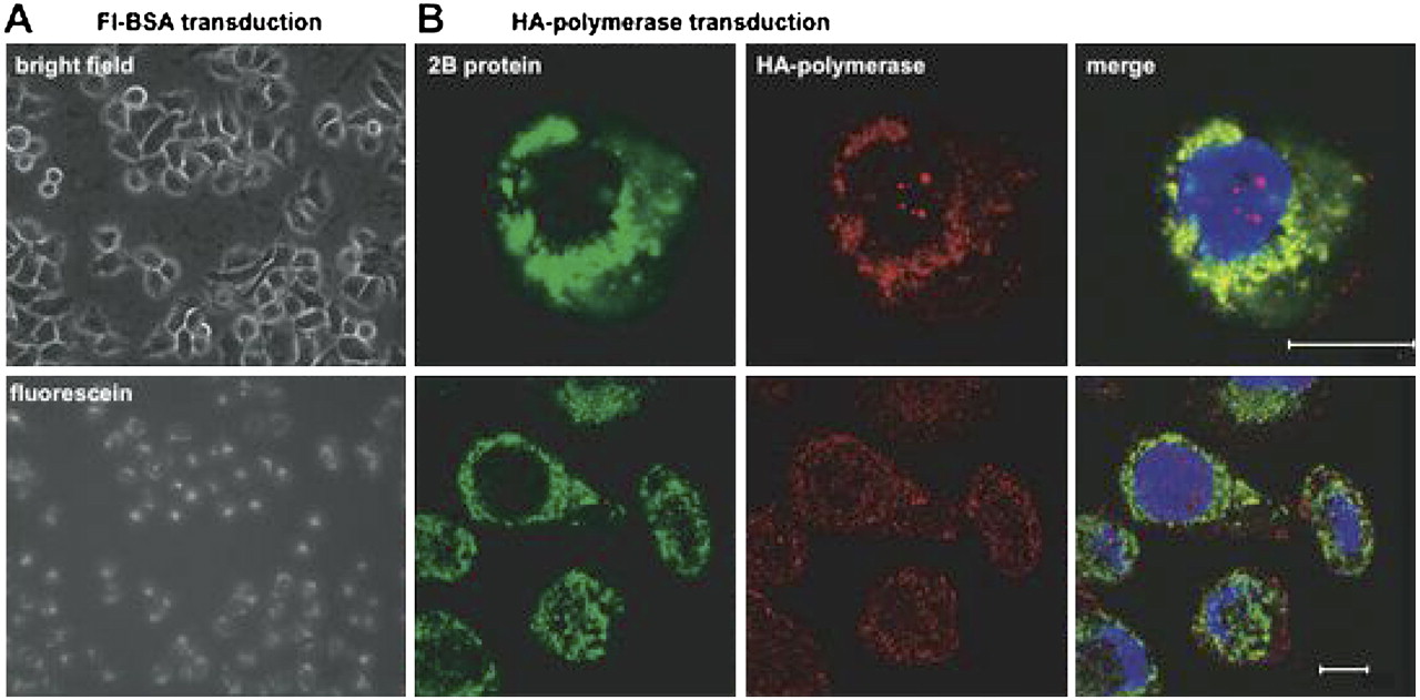
FIGURE 4.
Visualization of transduced proteins in HeLa cells. (A) Epifluorescent light microscopy of HeLa cells transduced with BodipyFL-conjugated BSA. (Top) Visualization of a field of transduced cells under phase contrast; (bottom) visualization of the same field under a fluorescein filter set after the addition of trypan blue to quench extracellular fluorescence. (B) Confocal micrographs from V391L-infected, HA-polymerase-transduced HeLa cells. Visualization of the poliovirus membrane-associated RNA replication protein 2B (green), transduced HA-tagged 3D polymerase (red), and merged fields are shown. Scale bar, 10 microns.




