


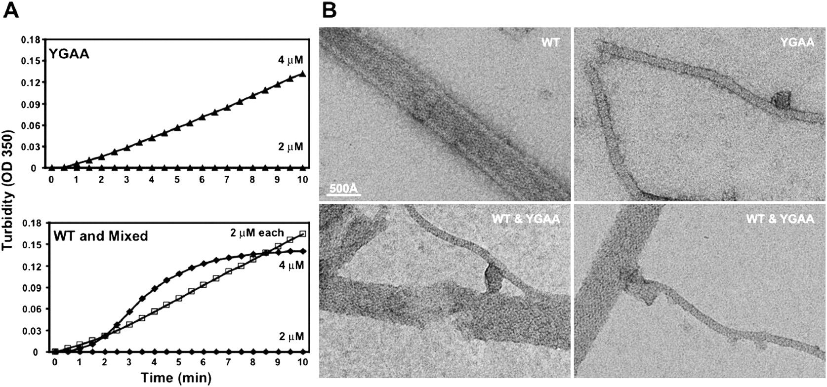
Formation and visualization of mixed poliovirus 3D polymerase oligomers. (A) Concentrated YGAA mutant poliovirus polymerase (top) or wild-type poliovirus polymerase (bottom) were diluted to 2 μM or 4 μM as indicated, and turbidity was monitored. A mixture of 2 μM of each polymerase was also monitored for the ability to oligomerize (bottom, □). (B) Negative stained electron micrographs of 1 μM purified wild-type polymerase (top, left), 1 μM purified YGAA mutant polymerase (top, right), and mixtures of 1 μM each (bottom). WT tubes are stain-filled, as seen by the high-protein density visible at the edges of the tube and the lower density in the tube center (protein is white). YGAA tubes are stain-excluding, suggesting tubes without a hollow center. Mixed samples show transition regions, as for example, the YGAA-like tube (bottom, left) that extends from a WT-like region of a thick tube. Scale bar, 500 Å.




