


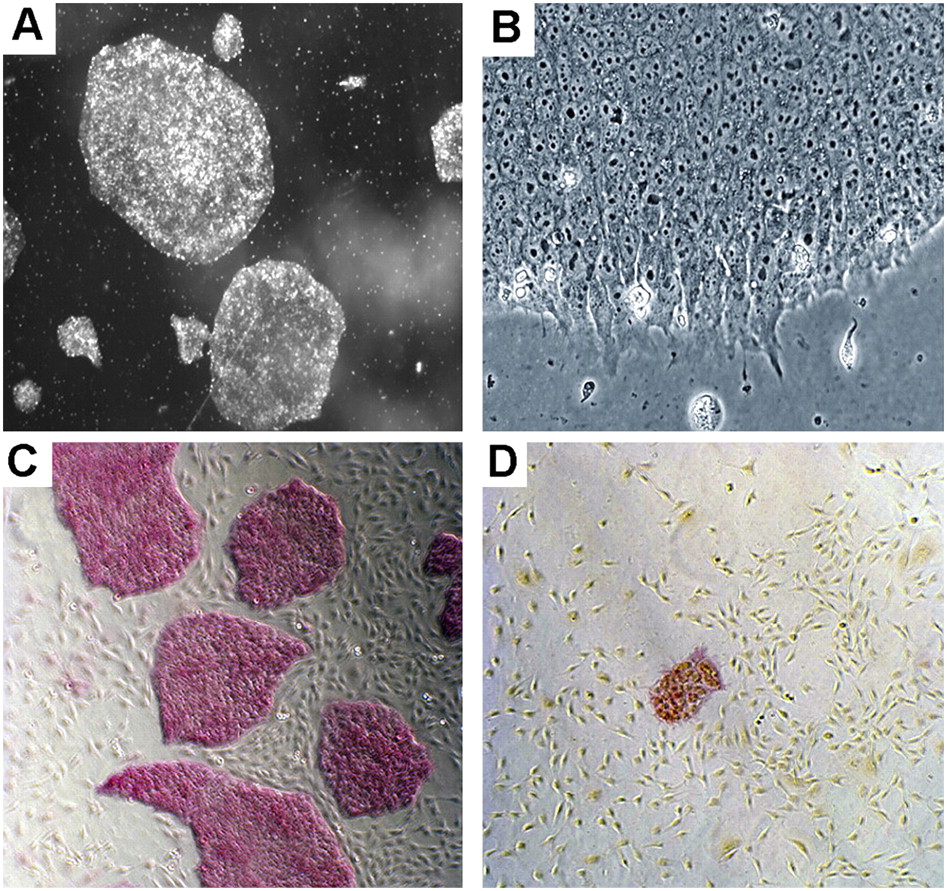
FIGURE 1.
Colony and cell morphology of hESCs grown under feeder-free, serum-free, and components-defined conditions. (A,B) Images of cell colonies taken at 20× and 200× magnifications, respectively. (C,D) Images of cell colonies resulted from cloning assays using our method and the traditional method, respectively. The pink color indicates undifferentiated alkaline phosphatase (AP) positive cells. Images were taken at a 40× magnification.




