


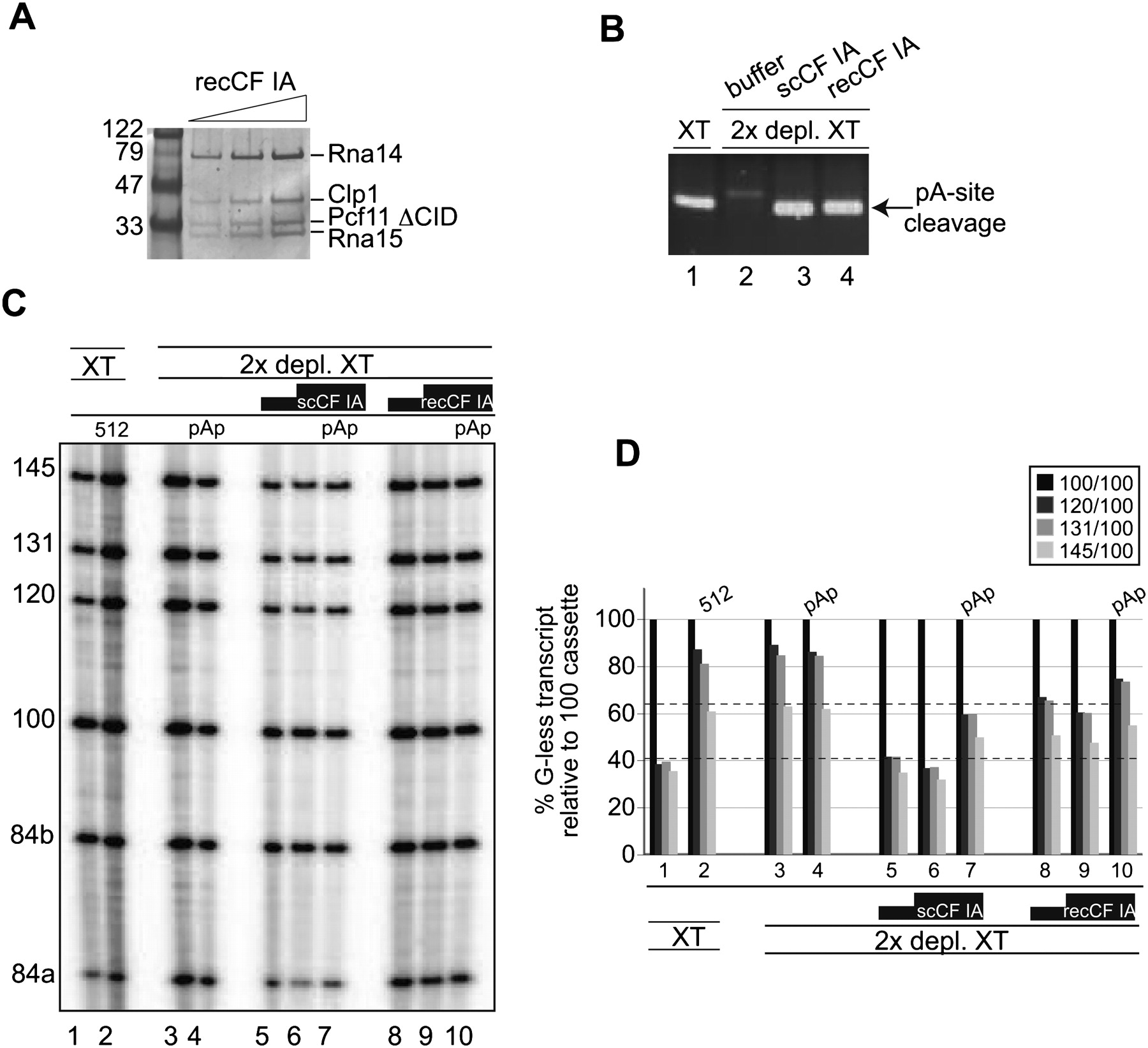
In vitro transcription/processing with recombinant CF IA factor (recCF IA. (A) Increasing amounts of recCF IA reconstituted from Pcf11ΔN288 (Pcf11 ΔCID), Clp1, Rna14, and Rna15 proteins were analyzed by SDS-PAGE and colloidal Coomassie G-250 staining. Molecular weight standards and their size in kDa are shown on the left. (B) 3′ end analysis as outlined in Figure 3D, of in vitro transcription/processing reactions with ProtA-tagged Rna15 extracts before (lane 1) and after (lanes 2–4) IgG-Sepharose depletion as described in Figure 3A. Reactions were complemented with CF IA factor purified from yeast extract (lane 3) or with reconstituted CF IA factor (lane 4). (C) In vitro transcription/processing reactions with constructs carrying the wild-type CYC1 terminator and with extracts before (lanes 1,2) and after (lanes 3–10) depletion of ProtA-tagged Rna15 protein. Two and four microliters of scCF IA and recCF IA factor, respectively, was added to depleted extracts as indicated. For control, reactions were also done with nondepleted extracts on templates carrying the mutant cyc1-512 terminator (lane 2) and in the presence of 4 mM pAp (lanes 4,7,10). (D) Quantification of results obtained in C.




