


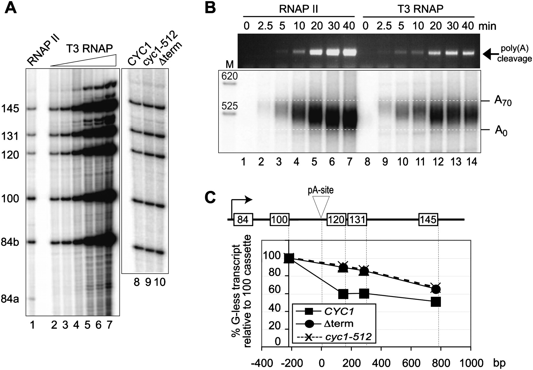
In vitro transcription/processing with T3 RNA polymerase. (A) In vitro transcription/processing reactions with wild-type extracts and templates carrying the CYC1 terminator driven by endogenous RNAP II (lane 1) or with increasing amounts of added T3 RNAP (lanes 2–7). The efficiency of termination/3′ end formation in T3 RNAP–dependent reactions was compared on templates carrying wild-type CYC1 (lane 8), mutant cyc1-512 (lane 9), or no terminator (lane 10). (B) 3′ end analysis as outlined in Figure 3D, of time-course of in vitro transcription/processing reactions dependent on extract endogenous RNAP II and T3 RNAP, as indicated. The upper panel shows nonradioactive PCR reactions that were separated on 1% agarose gels. The lower panel shows reactions where PCR was performed in the presence of 5′-32P-labelled primer B. Those reaction products were precipitated and resolved on denaturing 8.3 M urea/6% polyacrylamide gels. The approximate migration of 3′ ends carrying no or 70 adenosine residues, respectively, are indicated. (C) Quantification of results obtained in A.




