


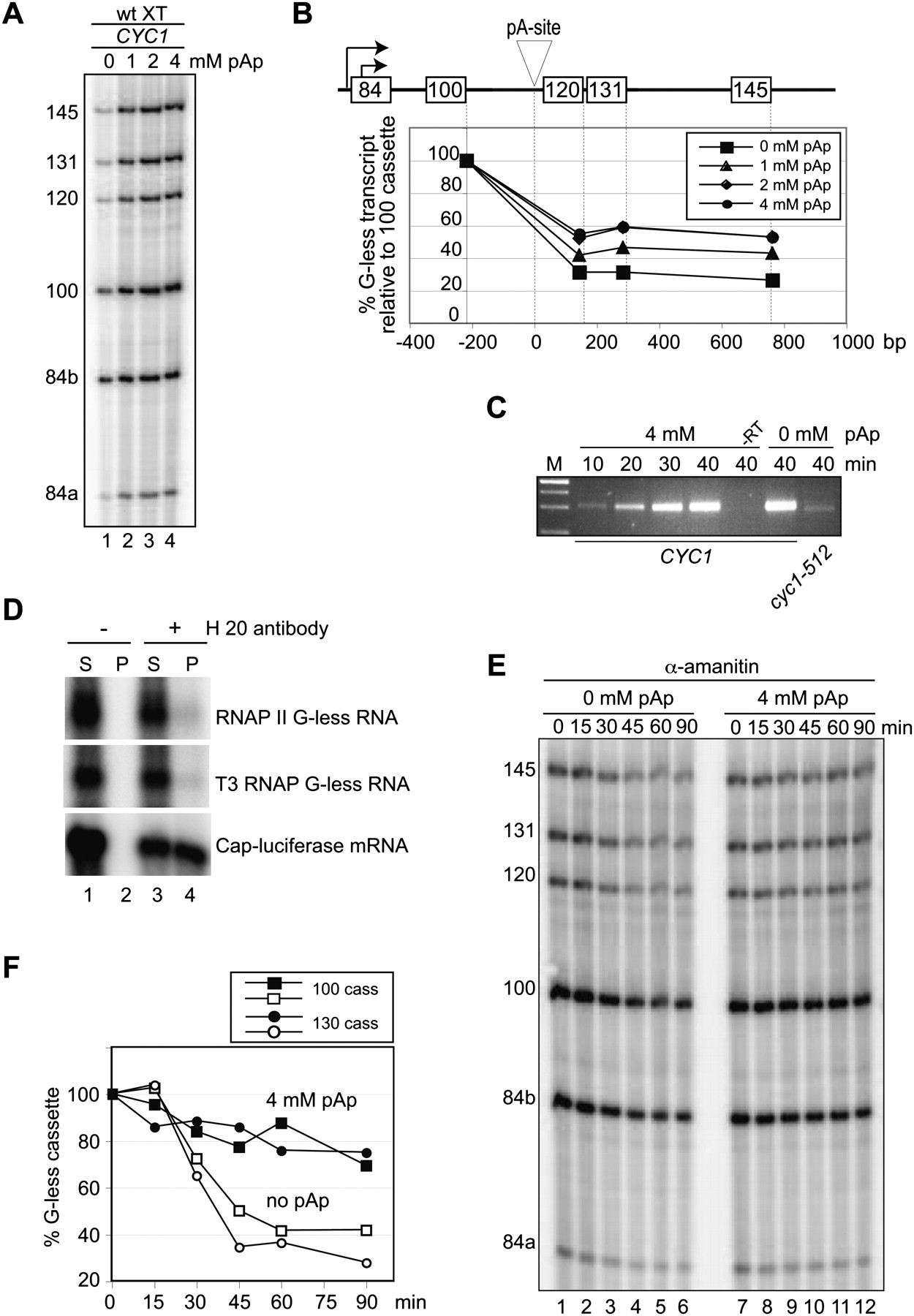
Effects of 3′,5′ adenosine bisphosphate (pAp) on in vitro transcription/processing with wild-type extracts. (A) In vitro transcription/processing with wild-type extracts with template carrying wild-type CYC1 terminator in the presence of increasing amounts of pAp, as indicated. (B) Quantification of results obtained in A. (C) 3′ end analysis as outlined in Figure 3D, of time-course of in vitro transcription/processing reactions in the absence and presence of 4 mM pAp. For control, a reaction lacking reverse transcriptase (−RT) was included for the 40-min time point, and the mutant cyc1-512 terminator was analyzed in the absence of pAp. (D) Immunoprecipitation of RNAs obtained from RNAP II and T3 RNAP-dependent transcription reactions, respectively. Luciferase mRNA served as positive control and was capped using the T7 Ribomax kit from Promega. Note that in vitro capping of the later RNA is not complete, accounting for the nonbound luciferase mRNA that is found in the supernatant (lane 3). RNAs bound to protein A–Sepharose in the presence and absence of anti-m7G antibody H 20 were separated into unbound (S) and bound fractions (P) and analyzed as described in the legend of Figure 1B. (E) Decay of G-less RNAs was analyzed following the in vitro transcription of the CYC1 terminator template with wild-type extracts. After 30 min of reaction time, RNAP II was inhibited by adding 20 ng/μL α-amanitin together with H2O (lanes 1–6) or 4 mM pAp (lanes 7–12). Aliquots were removed from the reaction at the indicated times and analyzed as shown in Figure 1C. (F) Quantification of 100 and 130 G-less cassettes from E.




