


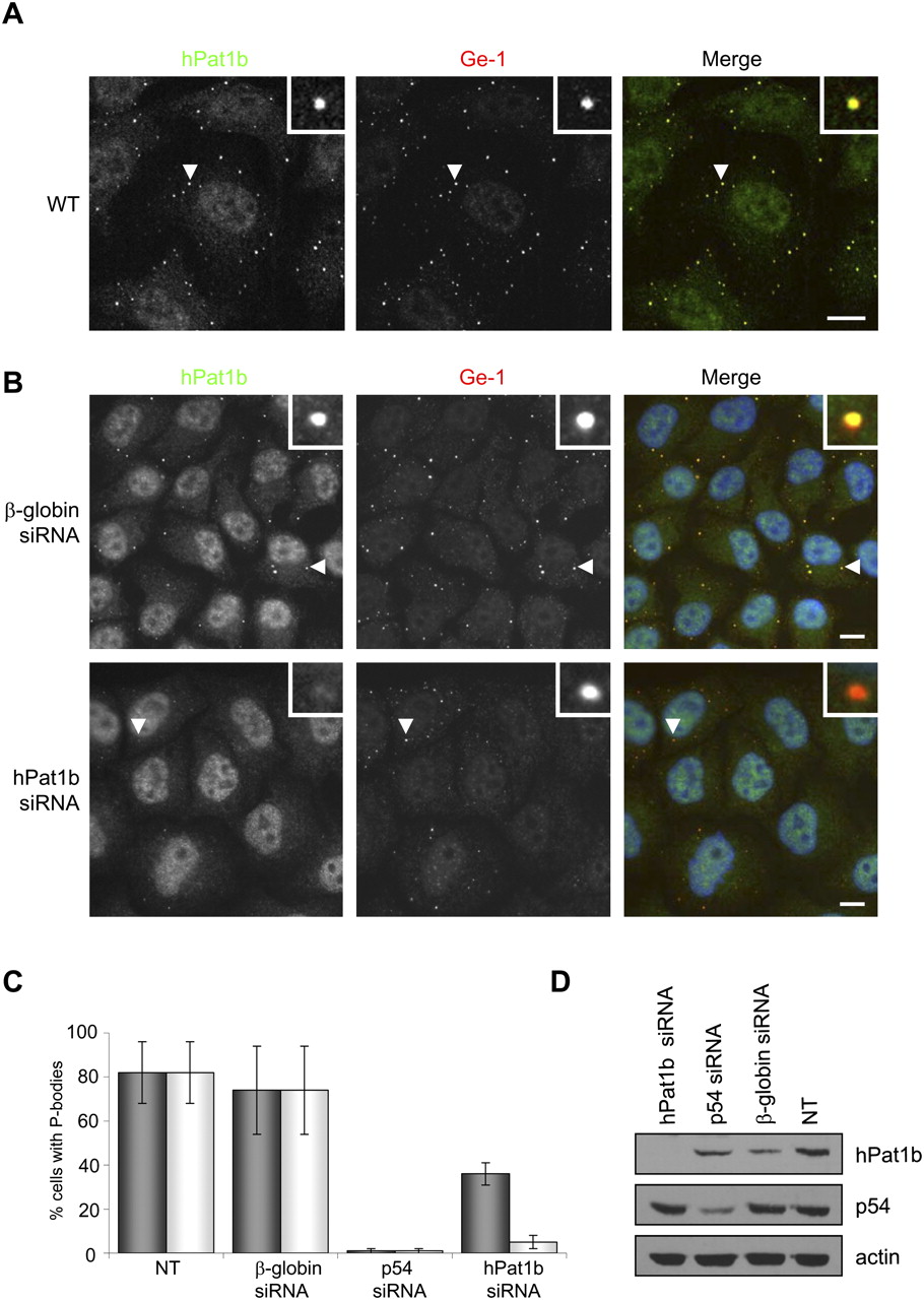
Endogenous hPat1b localizes to P-bodies and is not absolutely required for their formation. (A) Confocal imaging of HeLa cells stained with Pat1b antibody, with Ge-1 as a P-body marker. The white arrows point to the zoomed P-body (white box). Scale bar, 10 μm. (B) Depletion of hPat1b does not prevent P-body formation. hPat1b, p54, and β-globin siRNAs were transfected into HeLa cells, which were fixed and stained with hPat1b and Ge-1 antibodies 48 h post-transfection and visualized by fluorescence microscopy. The white arrows point to the zoomed P-body (white boxes). Scale bar, 10 μm. (C) Percentage of cells with P-bodies. The graph represents the percentage of cells with P-bodies as seen with either hPat1b (light gray) or Ge-1 (dark gray) antibodies, in nontransfected (NT) cells, or in cells transfected with control β-globin, p54, or hPat1b siRNA. Three independent experiments were performed and standard deviation bars are shown. (D) Western blot of the depletion effect of the different siRNAs used (30 μg protein loaded), with indicated antibodies.




