


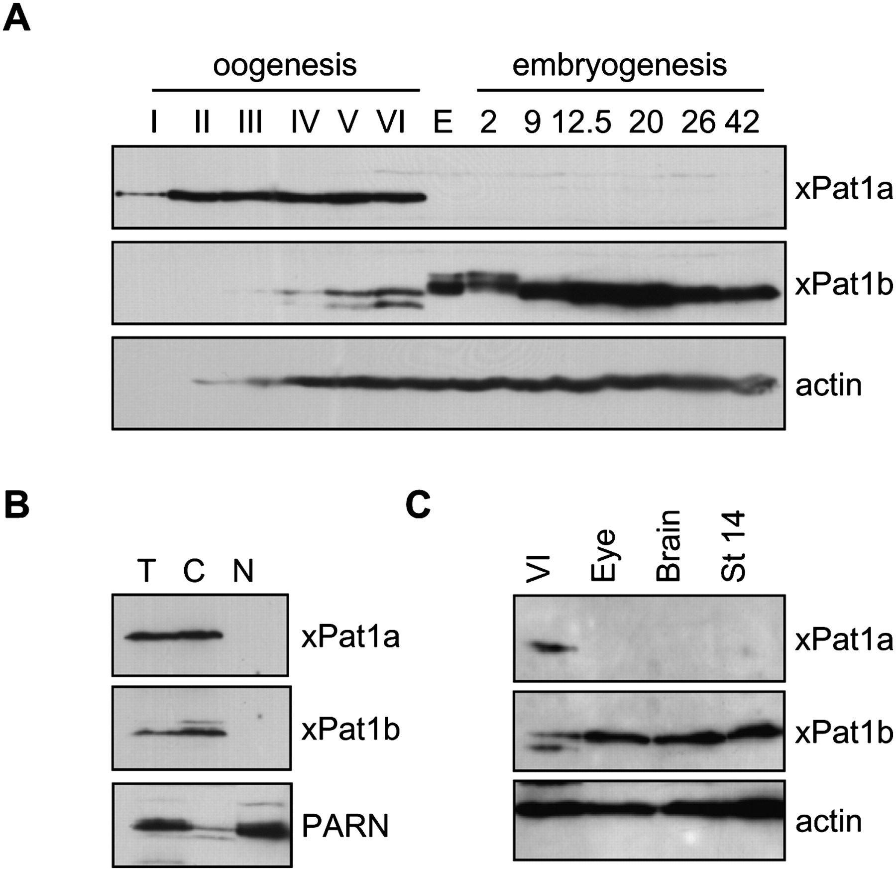
xPat1a and xPat1b expression profile in oogenesis and embryogenesis. (A) The expression levels of xPat1a and xPat1b were analyzed by Western blot using stage I–VI oocyte, egg, and different embryonic stages lysates (the stages of embryogenesis correspond to two-cell embryo; stage 9, midblastula; stage 12.5, gastrula; stage 20, neural fold closure; stage 26, tail-bud stage; stage 42, tadpole-like stage). Two cell equivalents were loaded. Actin was used as a loading control. (B) Both xPat1a and xPat1b are cytoplasmic proteins. Two cell equivalents of total (T), cytoplasmic (C), and nuclear (N) fractions from stage VI oocytes were analyzed by Western blot. PARN was used as nuclear control (Copeland and Wormington 2001). (C) xPat1b is expressed in eyes and brains of stage 42 embryos (tadpole-like stage). Actin was used as a loading control. One stage VI oocyte, 10 eyes, five brains of stage 42 embryos, and one stage 14 embryo were analyzed by Western blot.




