


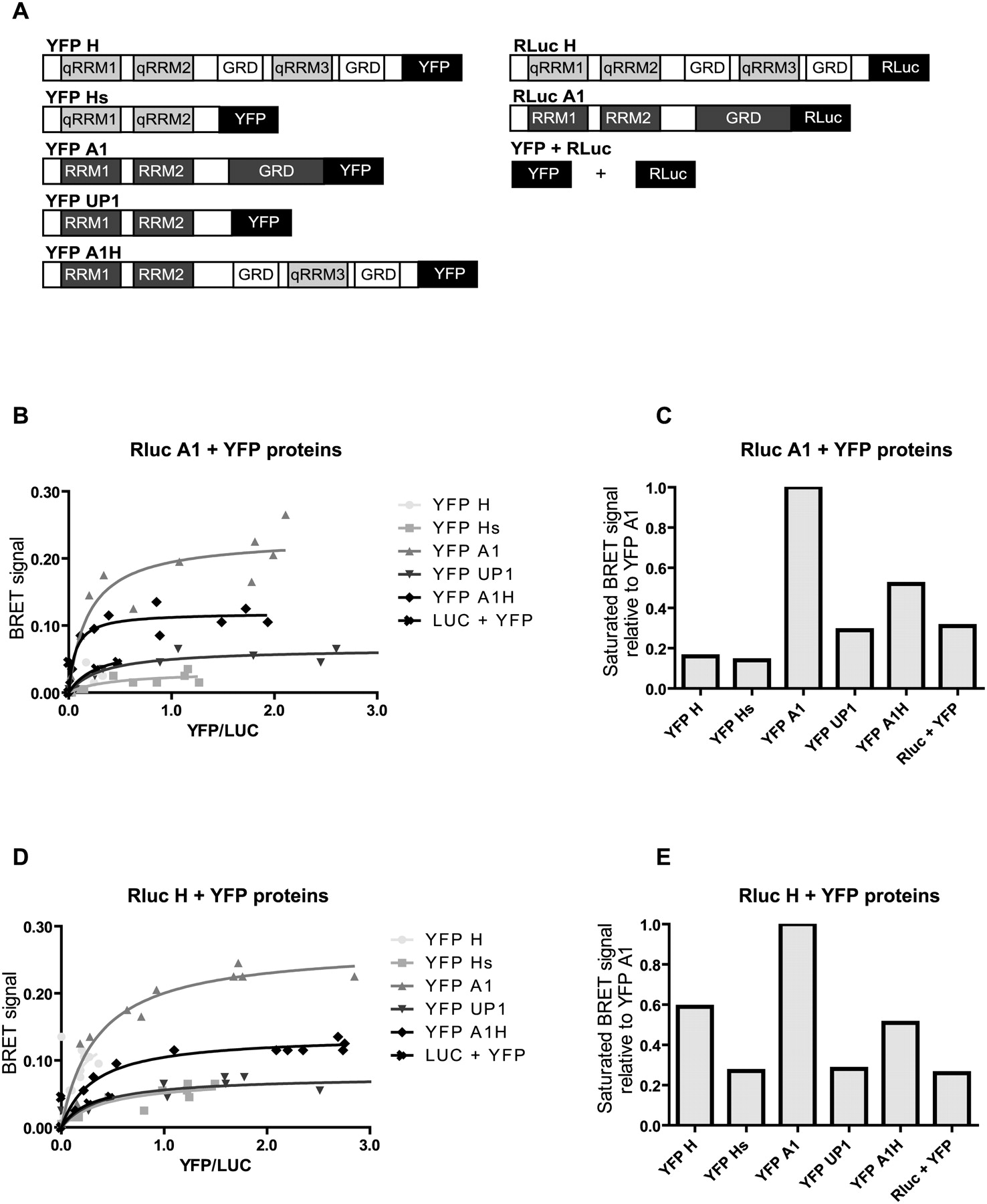
hnRNP H and hnRNP A1 can interact in live cells. (A) Structure of the YFP- and Rluc-tagged proteins used for BRET. (B, D) EcR 293 cells were transfected with 10 ng of plasmid expressing the Rluc version with increasing amounts (50, 100, 250, or 500 ng) of YFP-tagged plasmids. A control expressing only Rluc with an increasing amount of plasmids expressing only YFP was used (YFP + Rluc). Cells were collected 36 h post-transfection and the expression level of the YFP fused proteins was determined by fluorescence. The BRET signal was calculated as the fluorescence/luminescence after addition of 5 μM coelenterazine h (substrate for luciferase). The BRET signals were plotted relative to YFP/Rluc. (C, E) Plots showing the maximal saturated BRET signal for distinct combinations. The signals were normalized relative to YFP A1.




