


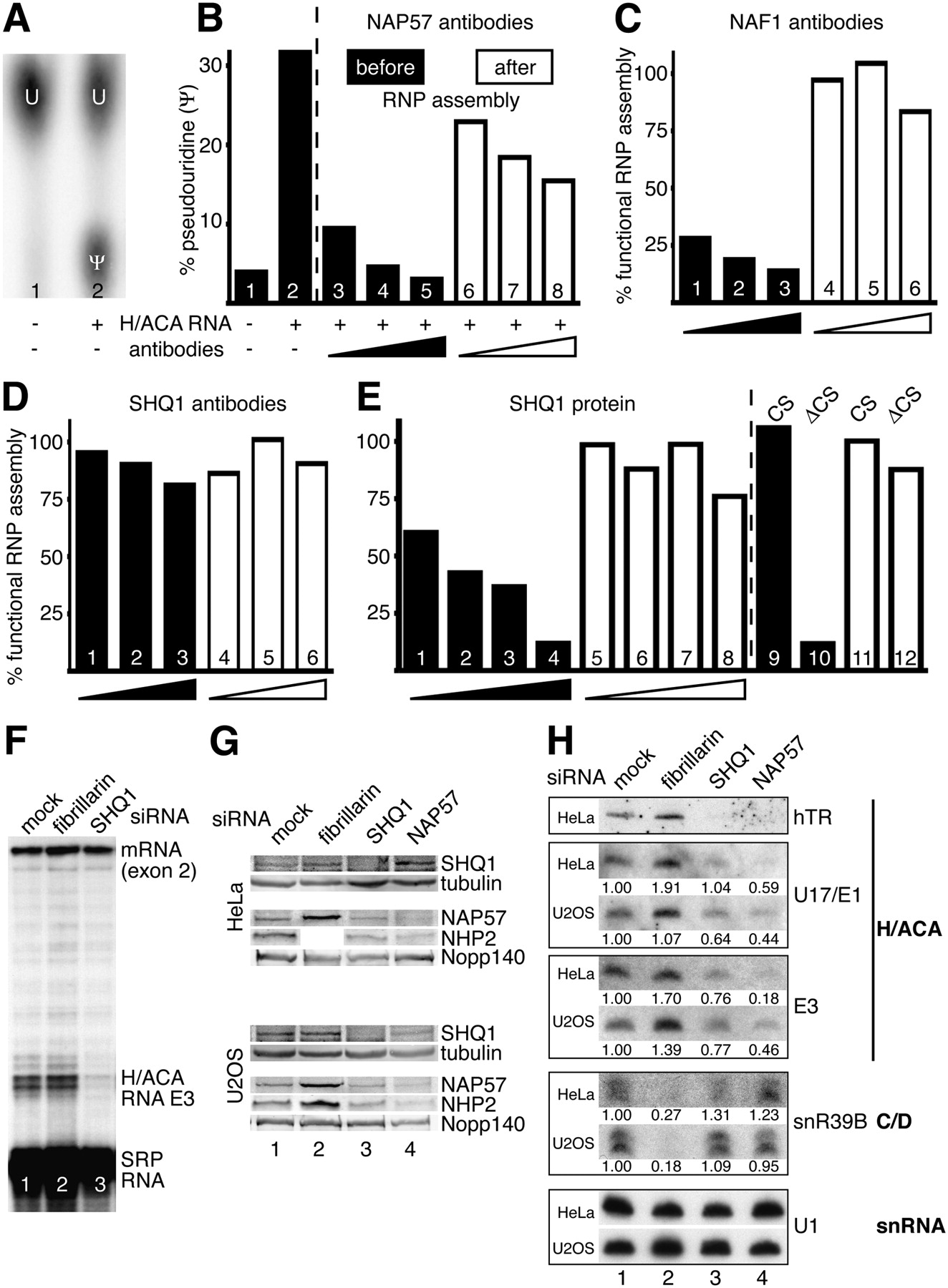
SHQ1 functions in H/ACA RNP assembly in vitro (A–E) and in vivo (F–H). (A) Autoradiograph of a thin layer chromatogram on which uridine (U) was separated from pseudouridine (Ψ) after liberation from a site-specifically 32P-labeled substrate RNA that was incubated in HeLa cytosolic S-100 extracts. Only when the cognate H/ACA RNA E3 was added to the extract for 30 min before substrate addition (lane 2), but not in its absence (lane 1), was the substrate RNA pseudouridylated, indicating that the in vitro transcribed H/ACA RNA was incorporated into a functional RNP during pre-incubation. Therefore, the pseudouridylase activity was used as a read-out for functional H/ACA RNP assembly. (B) Quantification of A (bars 1,2, respectively). Addition of increasing amounts of NAP57 antibodies prior to or concomitant with the H/ACA RNA (black bars) gradually inhibited activity (bars 3–6), indicating that they interfered with the incorporation of NAP57 into a functional RNP. Even if added after RNP assembly (open bars), the NAP57 antibodies were somewhat inhibitory. (C) Similarly, NAF1 antibodies only interfered with activity when added before (bars 1–3) but not after assembly (bars 3–6), showing that, as in vivo, functional assembly in this extract depended on NAF1. (D) SHQ1 antibodies did not interfere with activity whether added before (bars 1–3) or after assembly (bars 4–6). (E) However, addition of excess recombinant SHQ1 inhibited activity when added before (bars 1–4) but not after (bars 5–8) assembly, suggesting competition for a limiting factor, likely NAP57 itself. Indeed, this inhibitory activity resides in the NAP57-interacting SHQ1-ΔCS domain (bar 10) but not the SHQ1-CS domain (bar 9). Increasing amounts of antibodies were added as 1, 2, and 4 μL of the respective polyclonal anti-sera (B–D). Approximately 0.25, 0.5, 1.0, and 2.0 μg of recombinant proteins (Fig. 1B, lanes 1–3) were added (E). One hundred percent activity, i.e., functional assembly, corresponded to 34.6% (C), 31.0% (D), and 29.6% (E) of pseudouridine produced. The mean of duplicate values (B–D) and of one of several experiments (E) are shown. (F) Our inducible H/ACA RNA U2OS cell line (Fig. 3E) was treated for 3 d with siRNAs complementary to SHQ1 and the control nucleolar gene fibrillarin, induced for expression of the gene harboring the E3 H/ACA RNA in one of its intron, and the expression was analyzed using an RNase protection assay. Note, only in the case of SHQ1 knockdown (lane 3), but not that of fibrillarin (lane 2) or mock-treated cells (lane 1), did newly synthesized E3 fail to be stabilized, whereas the mRNA (exemplified by exon 2) was stably expressed in all three cases. Protection of SRP RNA with a separate probe served as a loading control. (G) Knockdown of the various proteins in HeLa (top) and U2OS cells (bottom) after treatment with siRNAs (targets indicated on top) was confirmed on Western blots with antibodies to the indicated antigens (right side). SHQ1 and the reference protein tubulin were extracted under low-salt conditions, whereas extraction of H/ACA RNPs (NAP57 and NHP2) and the nucleolar reference Nopp140 required 0.5 M salt. The antibodies were visualized using an infrared imaging system. Note in the case of fibrillarin knockdown, insufficient HeLa cells were harvested to analyze a separate gel for NHP2. (H) Autoradiographs of Northern blots of total RNA extracted from the siRNA treated cells in G and probed for the RNAs indicated on the right. The amounts of snoRNAs were quantified relative to those of U1 snRNA and expressed relative to those in mock-treated cells. The values for the H/ACA RNAs represent the means from two independent experiments. hTR could not be quantified because of interfering signal from spots, and levels of U17/E1 in HeLa cells appear too high due to the dark background. Finally, note that the H/ACA RNA E3 and the C/D RNA snR39B reside in different introns of the same gene.




