


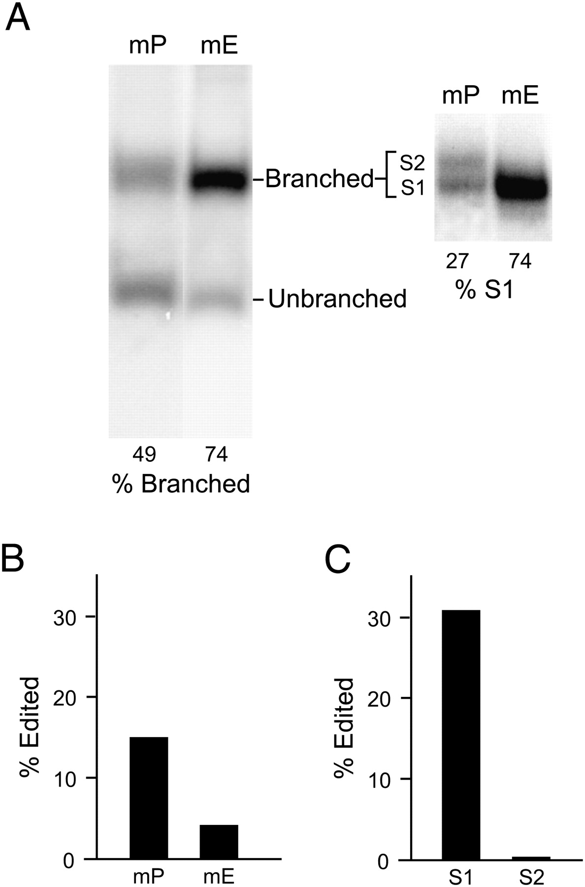
Comparison of the secondary structure distribution of Ecuadorian and Peruvian RNAs following transcription in vitro. (A) mP and mE RNAs were transcribed in vitro using T7 RNA polymerase and electrophoresed on a 6% nondenaturing polyacrylamide gel for 4 h at 4°C (left). The locations of branched and unbranched RNAs are indicated. Electrophoresis for 8 h at 4°C resolved two branched structures, S1 and S2, for mP RNA, but not for mE (Right). The migration of S1 was the same as the single branched species formed by mE RNA. (B) Editing of branched mP and mE RNAs. Branched mP and mE RNAs were gel-isolated and assayed for amber/W site editing as in Figure 1. (C) S1 and S2 mP RNAs were isolated from the gel in A and assayed for amber/W site editing as in Figure 1.




