


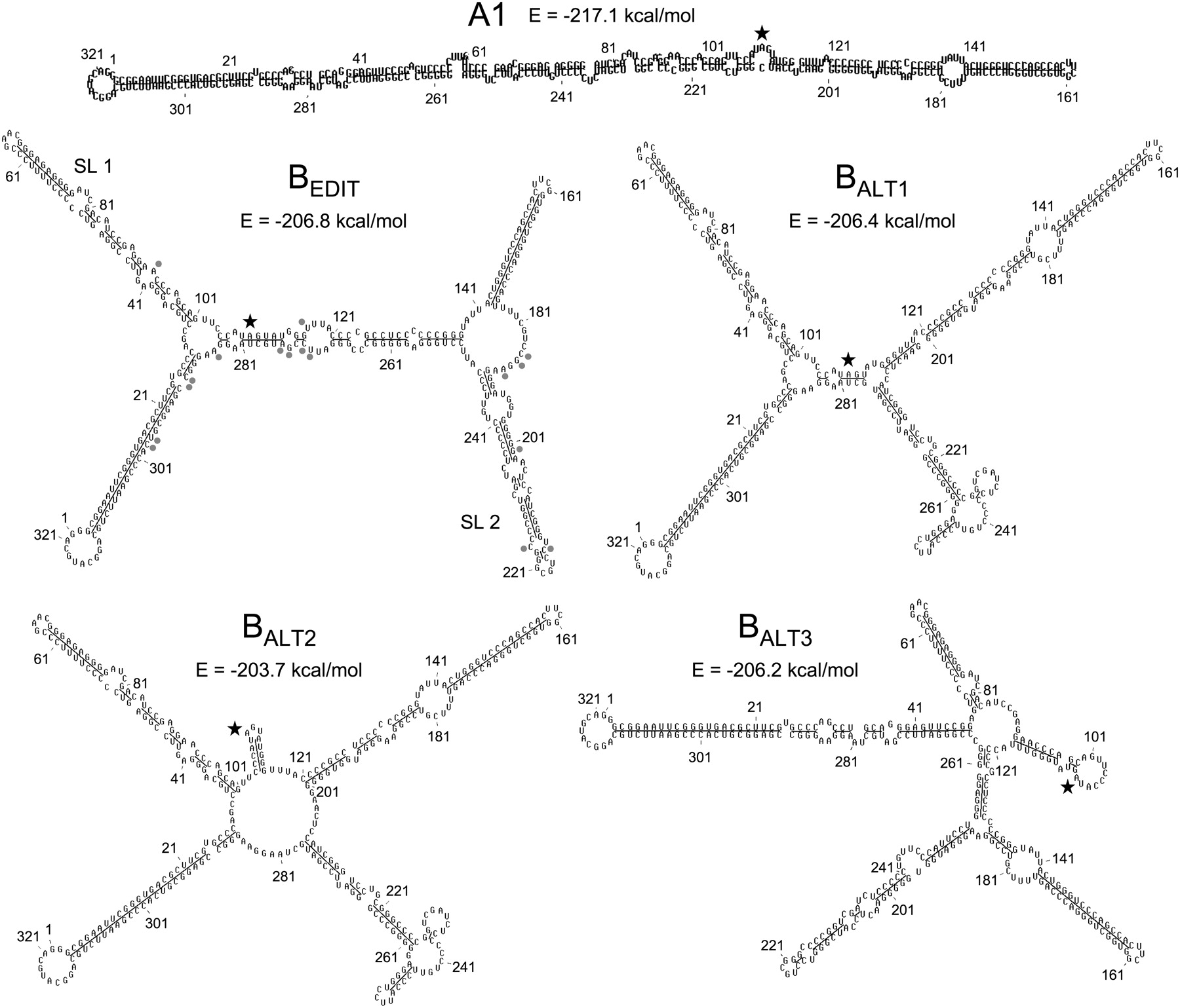
Secondary structures of the HDV-3 mP RNA predicted by MPGAfold. Black stars denote the location of the amber/W editing site in each predicted structure. The unbranched rod A1 is the best fit structure (lowest free energy structure achieved in MPGAfold runs). Structure BEDIT is very similar to that previously predicted for the Ecuadorian RNA mE (Linnstaedt et al. 2006) using efn2 energy rules. Extensions, or overruns, of the terminal stems in alternative branched structures BALT1, BALT2, and BALT3 alter base-pairing around the amber/W site and the lengths of stem–loops SL1 and SL2. Gray dots shown in the BEDIT structure denote locations of sequence differences between the mP and mE RNAs. Free energies predicted by MPGAfold are indicated. Energy calculations used the efn energy rules only, without the coaxial stem-stacking energy calculations for multibranch loops (efn2) (Mathews et al. 1999).




