


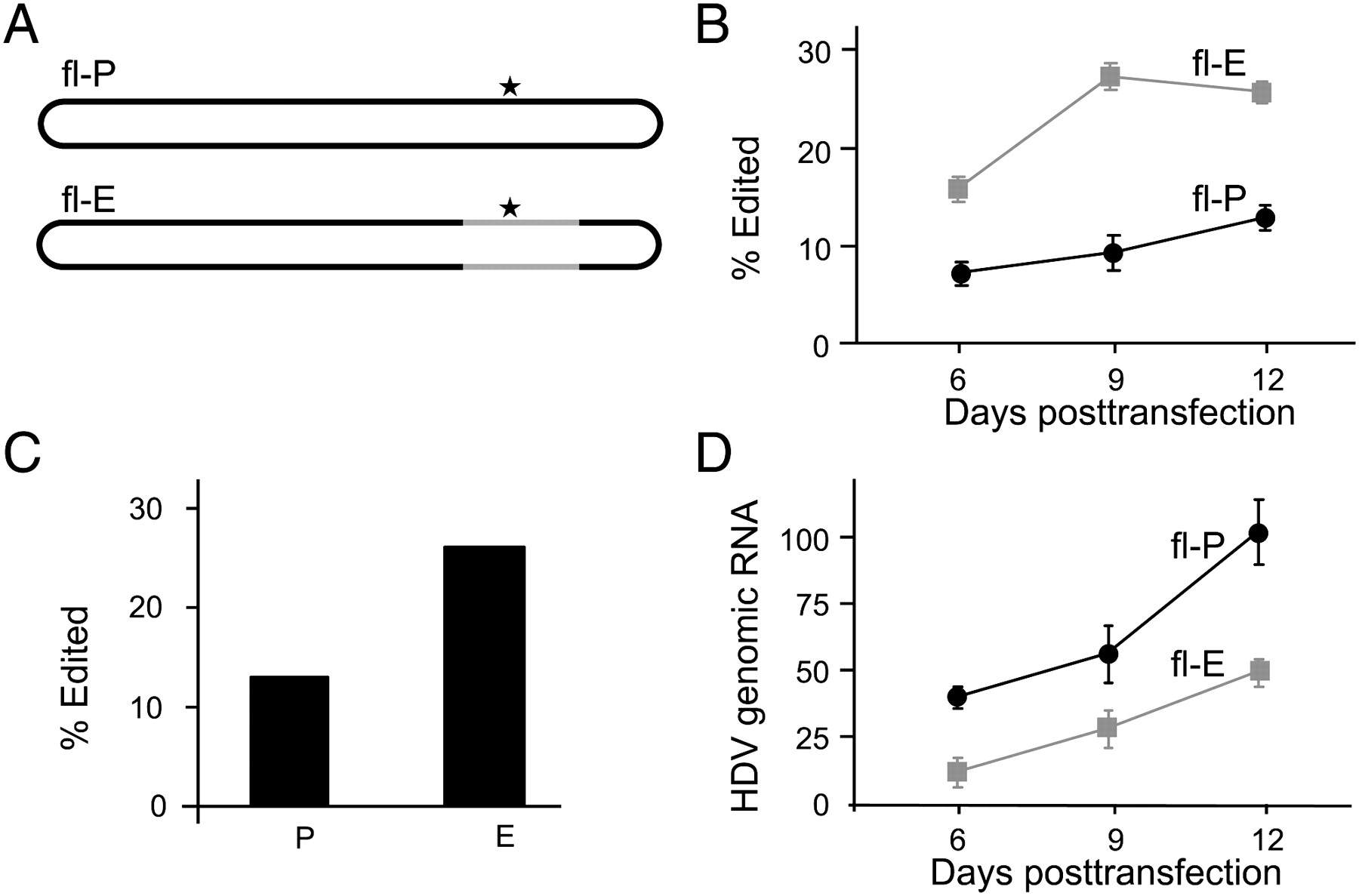
Comparison of editing in replicating RNAs. (A) Schematic diagram of replication-competent full-length RNAs fl-P and fl-E used to compare editing of replicating Peruvian and Ecuadorian HDV RNAs. The full-length Peruvian RNA, fl-P, is generated from a 1.2-length cDNA expression construct following transfection of Huh-7 cells (Casey and Gerin 1998). For the Ecuadorian RNA we created a chimeric construct to generate a full-length RNA, fl-E, that contains 15 of the Ecuadorian sequence differences in the branched structure that forms the substrate for editing (Fig. 1); the remainder consists of Peruvian sequences. Black indicates Peruvian sequences, gray indicates sequences (positions on replicating RNA) derived from the Ecuadorian isolate. (B) Huh-7 cells were transfected with expression constructs for fl-P and fl-E RNAs. Amber/W site editing was assayed on RNA isolated 6, 9, and 12 d post-transfection. Percent editing was determined as in Figure 1. Results are the average of three independent experiments. (C) Analysis of editing in HDV RNA isolated from patient sera. (D) HDV RNA levels in Huh-7 cells transfected with either fl-P (black circles) or fl-E (gray squares) antigenomic RNA expression constructs. RNAs were harvested on the indicated days post-transfection and analyzed by blot hybridization for levels of HDV genomic RNA. Three independent transfections were analyzed; error bars indicate standard deviations.




