


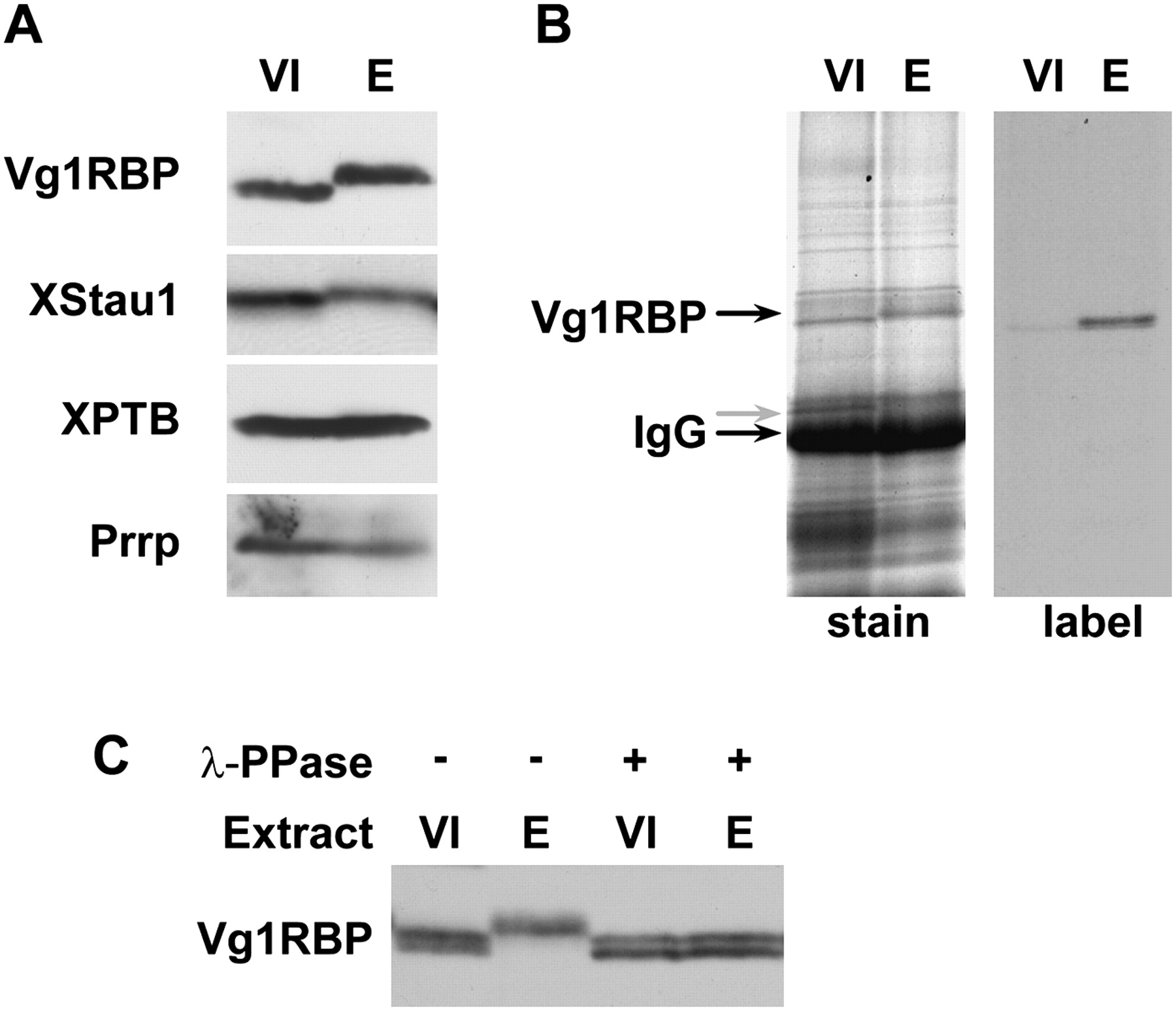
Vg1RBP is phosphorylated during meiotic maturation. (A) Vg1RBP undergoes an apparent shift in mobility during meiotic maturation. Protein samples from stage VI oocytes (VI) or progesterone-matured eggs (E) were analyzed by Western blotting using antisera directed against the proteins indicated on the left. (B) Vg1RBP is phosphorylated during meiotic maturation. Stage VI oocytes were incubated overnight in the presence (E) or absence (VI) of progesterone in a medium containing 33P orthophosphate. Vg1RBP was immunoprecipitated using anti-Vg1RBP antiserum and visualized either by autoradiography (right) or Coomassie Blue staining (left). Note that the overall immunoprecipitation efficiency is similar in both samples. The migration of Vg1RBP and of the IgG heavy chain is indicated. (C) The modification of Vg1RBP is phosphatase-sensitive. Protein samples from stage VI oocytes (VI) or progesterone-matured eggs (E) were treated with λ-phosphatase (λ-ppase) and compared to untreated samples using Western blot analysis. Note that under the PAGE conditions employed for this experiment, unmodified Vg1RBP migrates as doublet, reflecting the two allelic variants, A and D.




