


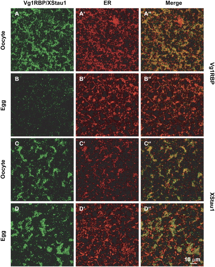
FIGURE 1.
Vg1RBP distribution in the vegetal subcortical region of oocytes and eggs. Confocal images of stage VI oocytes and progesterone-matured eggs stained with anti-Vg1RBP (A,B) and anti-XStau1 (C,D) antibodies. All samples were costained with anti-GRP94 antibodies to visualize the ER (A′–D′). (A″–D″) are the corresponding overlays of Vg1RBP/XStau1 (green) and GRP94 (red). The confocal sections were taken about 5 μm below the vegetal surface in a layer containing distinctive patches containing Vg1RBP and XStau1. Scale bar 10 μm.




