


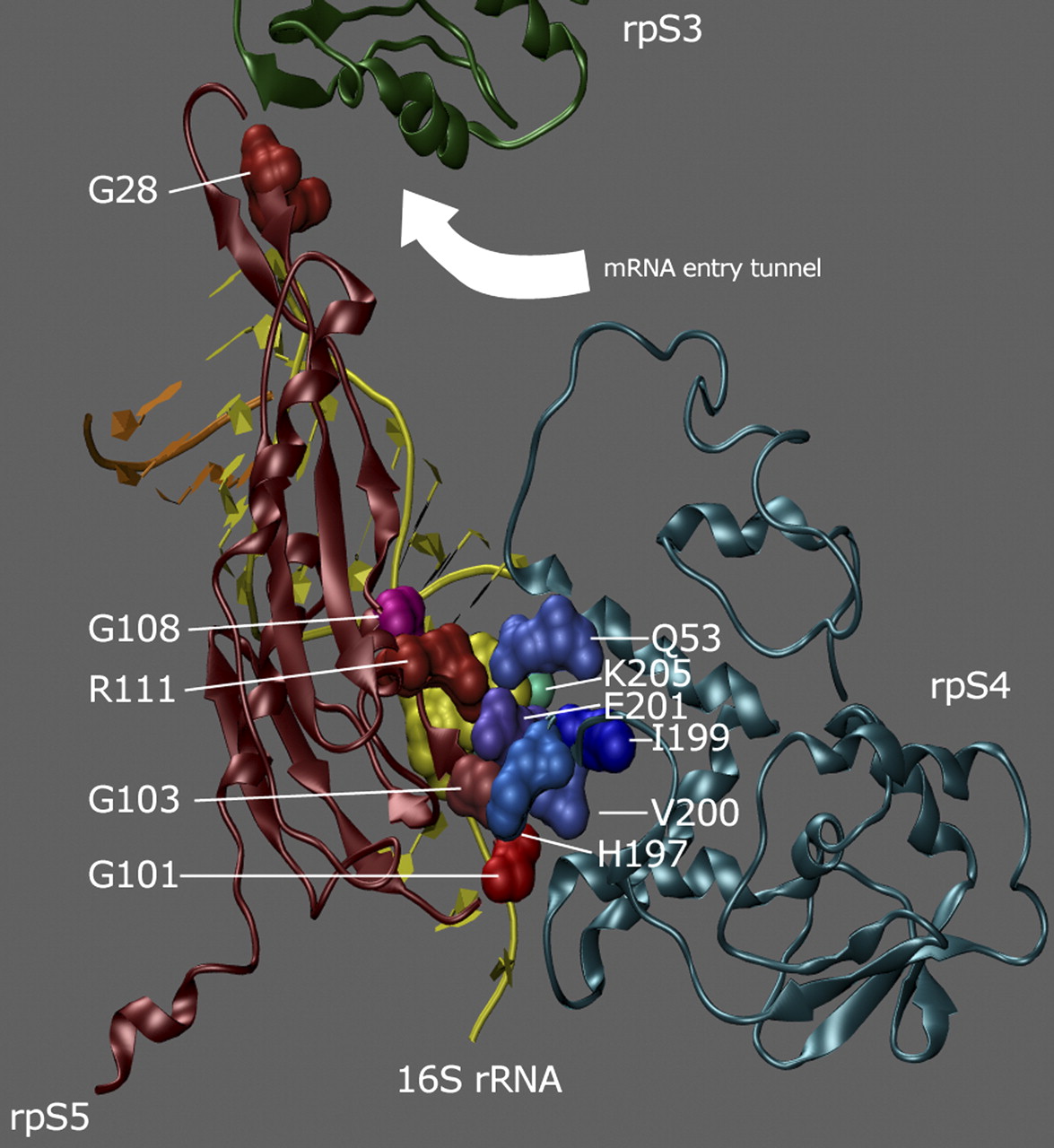
FIGURE 1.
Interactions at the rpS4•rpS5 protein–protein interface. Location of mutant residues analyzed in this work. The E. coli rpS4 (blue) and rpS5 (red) proteins and residues 5–25 of 16S rRNA are shown in cartoon form (PDB 2AVY, visualized using Visual Molecular Dynamics and MegaPOV); along with rpS3 (green cartoon), they form the ribosome helicase center through which the mRNA enters the ribosome (arrow). The mutant residues (labeled in white) are shown in space-filling format in shades of green for rpS4 and red for rpS5.




