


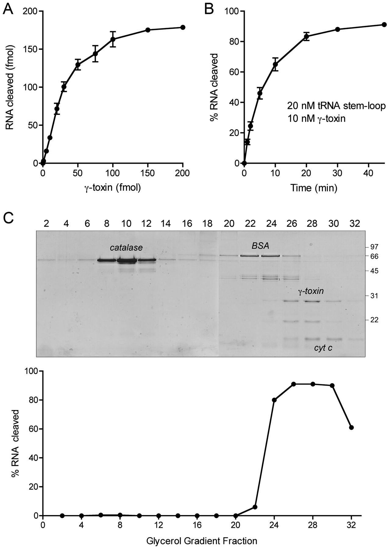
γ-toxin catalyzes multiple turnovers and sediments as a monomer. (A) Reaction mixtures (10 μL) containing 20 mM Tris-HCl (pH 7.5), 2 M TMAO, 200 fmol 32P-labeled 17-mer RNA, and increasing amounts of γ-toxin as specified were incubated at 4°C for 30 min. The extents of RNA cleavage are plotted as a function of input γ-toxin. Each datum is an average of three separate titration experiments ± SEM. (B) A reaction mixture (90 μL) containing 20 mM Tris-HCl (pH 7.5), 2 M TMAO, and 20 nM 32P-labeled 17-mer RNA, and 10 nM γ-toxin was incubated at 4°C. Aliquots (10 μL) were withdrawn at the times specified and quenched with formamide-EDTA. The extents of RNA cleavage are plotted as a function of time. Each datum is an average of three separate experiments ± SEM. (C) Aliquots (15 μL) of even-numbered glycerol gradient fractions were analyzed by SDS-PAGE (top). The Coomassie-blue stained gel is shown. The γ-toxin, catalase, BSA, and cytochrome c polypeptides are indicated. Molecular weight calibration for the PAGE analysis (kDa) is indicated at right. Reaction mixtures (10 μL) containing 20 mM Tris-HCl (pH 7.5), 2 M TMAO, 20 nM 32P-labeled 17-mer RNA, and 1 μL of the even-numbered glycerol gradient fractions were incubated at 4°C for 10 min (bottom). The extents of RNA cleavage are plotted.




