


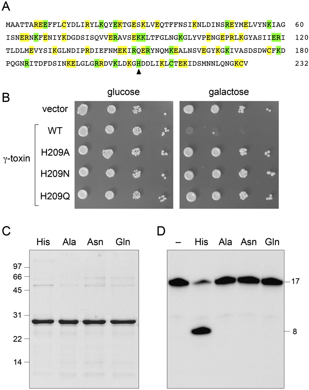
Mutational analysis of γ-toxin and essential catalytic role of His209. (A) The amino acid sequence of the intracellular version of K. lactis γ-toxin used in this study is shown. The amino acids subjected to alanine scanning are highlighted in green (essential residues) and yellow (nonessential residues). The lone histidine is denoted by an arrowhead. (B) Aliquots of serial dilutions of S. cerevisiae cells with a plasmid-borne galactose-regulated wild-type (WT) γ-toxin gene or H209A, H209N, or H209Q mutants thereof were spotted on glucose- or galactose-containing agar medium, in parallel with cells bearing an empty plasmid vector. The plates were photographed after incubation at 30°C for 2 d (glucose) or 3 d (galactose). (C) Aliquots (3 μg) of the recombinant tag-free γ-toxin C13A-C177A-C231A polypeptides were analyzed by SDS-PAGE. The Coomassie-blue-stained gel is shown. The amino acid at position 209 is indicated above each lane. The positions and sizes (in kDa) of marker polypeptides are indicated on the left. (D) Anticodon nuclease reaction mixtures (10 μL) containing 20 mM Tris-HCl (pH 7.5), 2 M TMAO, 200 fmol 5′ 32P-labeled 17-mer stem–loop RNA (see Fig. 2D), and 200 fmol of the indicated γ-toxin preparations were incubated at 4°C for 30 min. Enzyme was omitted from the control reaction in lane “−.” The products were analyzed by denaturing PAGE. An autoradiograph of the gel is shown, with the sizes (in nucleotides) of the substrate and product indicated on the right.




