


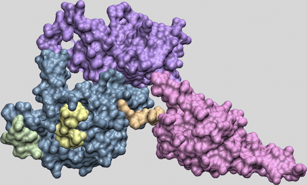
FIGURE 1.
Structure of human eRF1. Domain 1 (blue), domain 2 (red), and domain 3 (violet) of human eRF1 are represented in a van der Waals surface format. The two conserved motifs in domain 1 are shown in contrasting colors: TASNIKS, green; YxCxxxF, yellow. The linker between domain 1 and domain 2 (orange) is the site of the fusion of the E. octocarinatus or T. thermophila domain 1 with domain 2 and 3 of the S. cerevisiae enzyme.




