


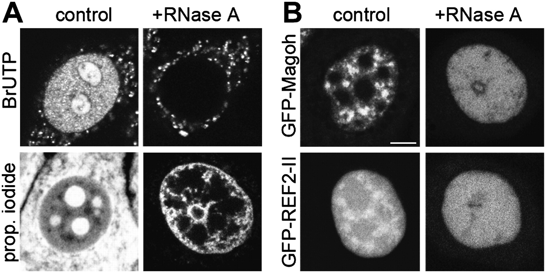
Accumulation of EJC in speckles is RNA dependent. The patterns of nascent RNA, nucleic acids, GFP-Magoh, and GFP–REF2-II were examined in intact cells subjected to RNA digestion. (A) Upper row: nascent RNA was labeled by BrUTP incorporation for 30 min. Cells were either fixed (left) or subsequently reinjected with RNase A (right) and then fixed. Control cells displayed the typical nascent RNA pattern, while in RNase A injected cells, no RNA was detected within the nuclei. Lower row: propidium iodide was used as a relocation marker for injected cells and to follow redistribution of nucleic acids upon RNase A injection. RNA digestion led to re-distribution of the propidium iodide signal and strong fluorescence appeared at the nuclear periphery in the nuclear center. (B) RNA digestion led to the disappearance of the typical speckled pattern of GFP–Magoh and GFP–REF2-II (examples show living/unfixed cells). Scale bar = 5 μm.




