


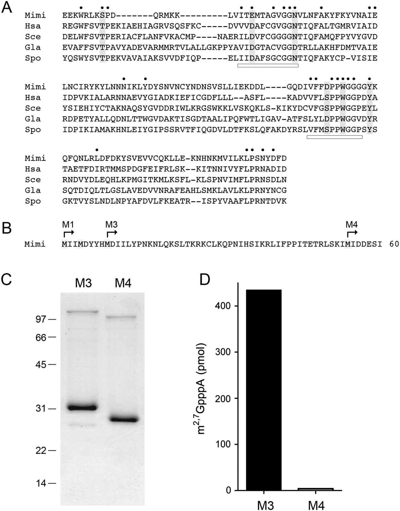
Mimivirus Tgs. (A) The amino acid sequence of MimiTgs (Mimi) from amino acids 91–229 is aligned to the sequences of the homologous cap guanine-N2 methyltransferases of Homo sapiens Tgs1 (Hsa), Saccharomyces cerevisiae Tgs1 (Sce), Giardia lambia Tgs2 (Gla), and Schizosaccharomyces pombe Tgs1 (Spo). Gaps are indicated by dashes. (●) Positions of amino acid side-chain identity/similarity in all five proteins. The AdoMet-binding motif and a putative m7G-binding motif are denoted by horizontal bars. Positions in MimiTgs that were targeted for mutational analysis in the present study are highlighted in shaded boxes. (B) The N-terminal 60 amino acids of the predicted MimiTgs polypeptide are shown, with methionines highlighted in shaded boxes. The translation start sites for the recombinant MimiTgs proteins M1, M3, and M4 are indicated by arrows. (C) MimiTgs purification. Aliquots (5 μg) of the purified tag-free M3 and M4 proteins were analyzed by SDS-PAGE. The Coomassie Blue-stained gel is shown. The positions and sizes (in kDa) of marker polypeptides are indicated on the left. (D) Methyltransferase reaction mixtures (10 μL) containing 1 mM m7GpppA, 50 μM [3H-CH3]AdoMet, and 0.5 μg of purified M3 or M4 protein were incubated for 15 min at 37°C. The products were analyzed by ascending TLC in 0.05 M (NH4)2SO4. The extents of 3H-methyl transfer to the cap dinucleotide are shown.




