


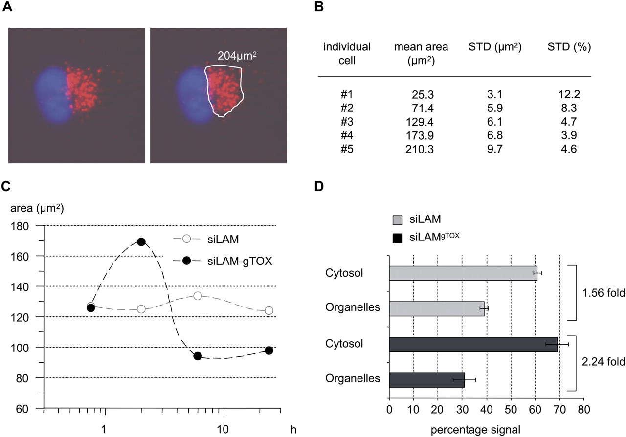
Fluorescence microscopy and cytosol/organelle separation indicate time-dependent release of siLAM-gTOX from perinuclear compartments. (A) Example for defining signal boundaries (white line) and for quantification of areas containing fluorescently labeled siRNA (red). Nuclei are stained with Hoechst 33342 and are shown in blue. (B) Area measurement of five individual cells differing in total fluorescent area. Determination of signal boundaries was repeated 10 times for each cell. Standard deviations of this area measurement are indicated as total area and as percentage of the mean. (C) The mean area of siRNA signals measured in ECV-304 cells (n = 40) at the indicated time points shows an initial increase in areas and a subsequent decrease for siLAM-gTOX but constant areas for siLAM. (D) The distribution of radiolabeled siLAM or siLAM-gTOX in organelles and cytosolic cell fraction shows increased cytosolic availability of the siLAM–peptide conjugate versus siLAM. Radioactively labeled siRNA was delivered by PS stimulation in ECV-304 cells. Radioactive signals in cytosolic and organelle fractions were quantified by liquid scintillation counting. The total radioactivity was set to 100%. For siLAM-gTOX the cytosolic amount was 2.24-fold greater than the organelle-associated amount versus a factor of 1.56 observed for siLAM. (g) Guide strand; (TOX) TQIENLKEKG.




