


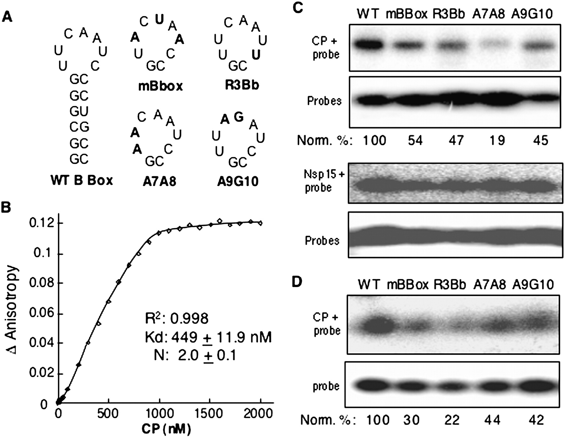
Protein–RNA binding assay in vitro. (A) Structure and sequence of WT B box and the mutants. The RNA structure was modeled with the Mfold program (Zuker 2003). Substitutions in the loop sequence are shown in bold, and the names are shown below the RNA structure. (B) Determination of the CP–RNA binding affinity by anisotropy experiment with fluorescence-labeled WT B box RNA. The fluorescein-labeled RNA was at 0.2 μM in a buffer containing 50 mM Tris (pH 7.5) and 50 mM NaCl; BMV capsid protein was added to a final cumulative volume of less than 5% of the sample. All data were corrected for the background intensity of the buffer. Binding data were fitted into the Hill Equation using KaleidaGraph software (Synergy Software). (C) UV–cross-linking assay. About 100 ng of CP was UV cross-linked with radioactively labeled WT B Box or mutants as described in Material and Methods. “Probes” denotes the labeled RNAs used in the cross-linking assay. (D) Electrophoretic mobility shift experiment of BMV CP and RNAs. The radioactively labeled RNAs were incubated with 500 ng of purified CP at RT for 30 min and then subjected to electrophoresis in a native gradient gel of 3%–12% (Invitrogen) as described by the manufacturer. The quantification was normalized with the probes used in the experiment.




